There are no treatments for aortic stenosis, a heart valve problem afflicting as many as a fifth of people over 65. The only solution is to modify the affected valve by implanting a prosthetic device that’s suited to the anatomy and the pumping action of a patient’s heart. While most cases are straightforward, some are more complex. 3D printed hearts could help cardiologists gain greater insight into how a patient’s heart is pumping and find a better fit.
In a paper published Feb. 22 in Science Robotics, bioengineers led by a team from Massachusetts Institute of Technology described how they used cardiovascular data from individual patients to print soft models of their hearts, complete with the ability to pump just as their hearts did. “All hearts are different,” first author Luca Rosalia, a graduate student in the bioengineering lab of Dr. Ellen Roche, said in a press release. “There are massive variations, especially when patients are sick. The advantage of our system is that we can recreate not just the form of a patient’s heart but also its function in both physiology and disease.”
A condition called severe aortic stenosis, which warrants intervention, occurs when the width of the aortic valve drops below 1 square centimeter. Patients over 65 are typically treated with a minimally invasive procedure called transcatheter aortic valve replacement. In TAVR, cardiologists place one of two FDA-approved prosthetics—Medtronics’ Evolut R or Edwards Lifesciences’ SAPIEN 3—into the aortic valve. The prosthetic expands and integrates with the tissue, allowing the heart to pump blood effectively again.
The decision to perform TAVR is pretty straightforward in patients with the “classic” form of severe aortic stenosis, where the left ventricle ejection fraction is less than 50%, Nish Harshadkumar Patel, M.D., an interventional cardiologist at the Miami Cardiac & Vascular Institute of Baptist Health South Florida, who was not involved in the study, told Fierce Biotech Research. But it’s a more complex choice in patients with a form called paradoxical low-flow, low-grade severe aortic stenosis. In cases like this, “3D modeling would be very beneficial in understanding the biomechanical aspect of that valve and how it behaves in different flow dynamics,” he explained.
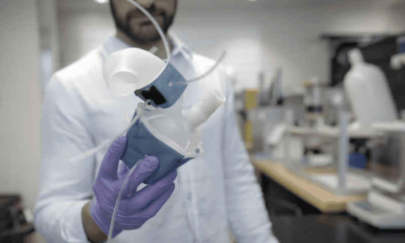
To see if they could offer a solution by way of 3D printed hearts, the Roche lab set out to create soft models that were anatomically and functionally representative of individual patients. They compiled CT data from 15 aortic stenosis patients and then reconstructed digital 3D renderings. These were subsequently used to print physical 3D models from an elastic plastic resin, the stretchiness of which mimics the heart muscle.
The team got the hearts to pump by wrapping them in sleeves similar to blood pressure cuffs. Each sleeve had four inflatable pockets that were designed to match the shape of the individual’s heart. The pace of contraction for each pocket could be adjusted individually, allowing the researchers to fine-tune the pumping motion so that it mirrored the patient’s real-life heart. A robotical air pump controlled the motion.
With the models built and pumping, the researchers took echocardiograms of the printed hearts in action and compared them to echocardiograms from the patients themselves. They found that the models recreated the pressure and blood flow seen in the patients, including those whose hearts had unique pumping mechanisms due to tissue remodeling caused by aortic stenosis.
Next, the researchers wanted to assess if they could also recreate the effect of TAVR on patients’ hearts. They implanted either the Evolut R or SAPIEN 3 prosthetics into the models, then recorded their pumping action and compared it to post-implantation results from the actual patients. Once again, the models mapped to the patients’ improved flow dynamics.
To round out their study, the team undertook a potential real-world use case for the models: fitting patient hearts with different sizes of prosthetics to assess potential problems from a mismatch. Measurements from the models showed that undersized implants would indeed cause the complications that come from pumping dynamics associated with a condition called paravalvular leak and regurgitation.
Eventually, the team hopes to see their models integrated into pre-implant preparation. Ideally, routine patient imaging could be used to print the models within a day, and clinicians could use the information to see which implant works best, paper co-author Christopher Nguyen said in the press release.
Besides assessing implant fit prior to TAVR, the devices could also be used to help medical device companies test and optimize their products for a wide range of clinical scenarios and make them more accessible to those who can’t currently use TAVR, the resarchers noted in their paper. In the clinic, they could also be used to make procedures safer in patients with complex anatomy.
As more prosthetics make their way to the market, like Abbott’s newly FDA-approved TAVR implant, Patel believes the need for 3D printed models like the one from MIT is poised to grow. He envisions a future where clinicians can demonstrate to patients how much improvement they’ll get from a particular valve before it’s implanted.“You can place a virtual valve and show them the difference they’re going to have in terms of the pressure gradient,” Patel said.

