Cancer cells multiply out of control because they’re able to escape a mechanism known as programed cell death. But scientists at the Massachusetts Institute of Technology (MIT) and King’s College London have uncovered key functions of another pathway with a long evolutionary history that the body uses to dispose of those rogue cells.
During this process, called cell extrusion, cells are squeezed out of the lining of tissues. The team, led by Nobel laureate H. Robert Horvitz, Ph.D., found that the process is triggered when cells fail to replicate their DNA during cell division, according to a study published in Nature.
Because this so-called replication stress predisposes cells to mutation and is therefore linked to precancerous and metastatic cells, the researchers suggested extrusion may serve as a basic mechanism to eliminate cancer cells.
“The discovery that extrusion is driven by a failure in DNA replication was unexpected and offers a new way to think about and possibly intervene in certain diseases, particularly cancer,” Horvitz said in a statement.
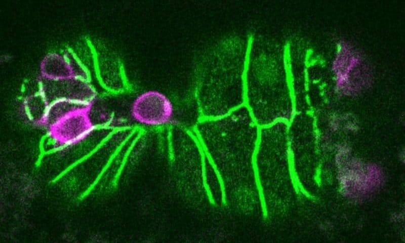
Horvitz shared 2002’s Nobel Prize in Physiology or Medicine for discovering and characterizing genes involved in cell death in Caenorhabditis elegans. And it was in the nematode worm that Horvitz’s lab first discovered cell extrusion as a backup mechanism to programmed cell death, or apoptosis.
For this study, Horvitz and colleagues set out to learn how extrusion is triggered. They knocked down over 11,000 C. elegans genes one by one to understand the role of the genes involved in the process. Many of the genes that they identified were active during the first steps of cell division, including DNA replication.
Further experiments showed the cells that were expelled appeared to get stuck in the DNA replication phase. These cells were very small compared to sister cells that divided from the same parent. They couldn’t complete the replication because of a lack of proteins and DNA building blocks, the team showed. One key component the cells lacked was an enzyme called LRR-1, which is critical for DNA replication. When this process hit a snag, a protein called CDK1 was inactivated through the ATR-CHK1 signaling pathway, leading to reduced cell adhesion and then extrusion, the researchers found.
The team then induced DNA replication stress in dog kidney cells. Doing so increased the rate of extrusion by more than threefold, the researchers reported. Inhibiting CHK1 with small-molecule chemicals suppressed the extrusion, indicating the process depends upon the ATR-CHK1 pathway.
In mammalian cells, the ATR-CHK1 pathway is known to activate p53, a transcription factor that suppresses cancer by promoting cell death. The MIT-led team showed that induced replication stress increased p53 levels in the dog cells. This suggests that in addition to promoting apoptosis, p53 may also play an important role in extrusion, the researchers said.
Many other scientific approaches are focused on stopping the uncontrolled growth of cancerous cells. A team at Vanderbilt University, for example, recently found that crippling a protein made by the gene TRAF3 could cause cells to divide without stopping.
The MIT team suggested their newfound understanding of extrusion could inform efforts to develop therapies aimed at stopping cancer cells early, when they encounter replication stress.
“Replication stress is one of the characteristic features of cells that are precancerous or cancerous,” Vivek Dwivedi, Ph.D., a post-doc at MIT and the study’s first author, said in the statement. “And what this finding suggests is that the extrusion of cells that are experiencing replication stress is potentially a tumor suppressor mechanism.”

