
Researchers at University of Tsukuba have achieved high-resolution visualization of cellular organelles, such as nuclei and mitochondria, using an external apodized phase contrast (ExAPC) microscope. By effectively suppressing halo artifacts—false images caused by light diffraction—the technique reveals the tightly regulated and remarkably stable movement, spatial arrangement, and morphology of these organelles. The team also observed unlabeled biomolecular condensate-like structures whose molecular components remain unidentified.
Cells contain various organelles, including the nucleus, which stores genetic information, and mitochondria, which generate the energy essential for biological processes. While these are “membranous organelles” enclosed by lipid bilayers, cells also contain “nonmembranous organelles” formed through molecular assembly without membrane encapsulation. The morphology, quantity, and spatial distribution of these organelles are crucial for maintaining cellular functions and determining cell fate.
Traditionally, fluorescence-based imaging has been the principal approach for visualizing organelles. However, this method has inherent limitations, including phototoxicity from intense illumination, photobleaching of fluorescent dyes, and restrictions on the number of structures that can be simultaneously observed.
To overcome these challenges, phase contrast microscopy—which enables the visualization of transparent structures without the need for staining—has regained attention. Yet, conventional phase contrast techniques are prone to halo artifacts caused by diffraction.
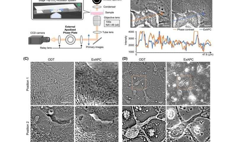
In their study published in The FEBS Journal, the researchers used ExAPC microscopy, which incorporates an optical mechanism to suppress halo formation. This enabled high-resolution, label-free observation of multiple organelles, including nuclei, nucleoli, and mitochondria, during dynamic cellular events such as the cell cycle.
Notably, for the first time, the researchers clearly visualized biomolecular condensate-like structures of unknown composition. Examination of lipid droplet growth, mitochondrial fission and fusion, and cellular responses to drugs revealed that while individual organelles display diverse behaviors (heterogeneity), the overall system maintains a high degree of order and stability (robustness).
These findings highlight ExAPC microscopy as a powerful tool for capturing biological dynamics in their native state. The technology holds significant potential for elucidating pathological conditions associated with altered organelle morphology, quantity, and spatial organization—such as cancer, metabolic disorders, and neurodegenerative diseases—and may contribute to the development of novel diagnostic and therapeutic strategies.