
Scientists have grown a scalable new embryo-like model that unshrouds some mysteries of early human development, including blood cell formation, or hematopoiesis—a first for the field.
In an article published Dec. 13 in Nature, a team of researchers from the University of Pittsburgh described how they created so-called heX-Embryoids, which are self-organizing structures smaller than the tip of a sewing needle. The models will be used to understand the first stages of blood cell development in humans, to perform toxicology studies and to figure out how to grow cells more effectively for therapeutic applications such as blood transfusions or cell therapies, among other potential uses.
“Our embryo-like model will unlock the ‘black-box’ of human development, which could help solve the mystery of why 60% of pregnancies fail in the first two weeks—before the mother even misses a menstrual period—and pave the way for new therapies,” Mo Ebrahimkhani, M.D., senior author, said in a press release.
The development is the second major triumph this year in the quest to design a model of the human embryo, a pursuit that has sparked both excitement and ethical concerns among the scientific community and the public. In September, researchers from the Weizmann Institute of Science in Israel published an article in Nature describing how they created a human embryo model that recapitulated the basic structure of real ones from implantation to around 14 days post-fertilization. That model even secreted hormones that turned pregnancy tests positive.
The Weizmann Institute’s development marked a turning point in the field, as it was the first time scientists had truly toed the line between embryo model and actual embryo, some unaffiliated researchers said. But the heX-Embryoids may ruffle fewer feathers: Unlike the earlier models, they don’t have the components necessary for forming a placenta, nor a closed yolk sac. Both features are necessary for implantation and growth into a “real” embryo.
“[heX-Embyroids] do not mirror the biology of the full integrated embryo and can provide a possibility for additional in vitro follow-ups with limited ethical concerns,” the researchers wrote in their paper.
The scientists grew the models from human adult skin cells that had been induced to revert to pluripotent stem cells, also known as iPSCs. The iPSCs’ genes were edited to express a pattern that could stimulate early tissue development when combined with the antibiotic doxycycline.
When the edited iPSCs were mixed with unedited iPSCs and doxycycline in a lab dish containing standard culture media—a departure from other methods that require complex concoctions of growth factors—they developed into an open yolk sac and embryonic tissue.
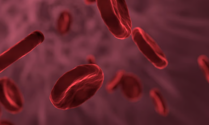
Unlike other embryo models, the structures also contained functional blood islands, or sites on the yolk sac where hematopoiesis takes place. Starting 16 days after the iPSCs were cultured, the blood islands began producing waves of precursor cells to bone marrow, red blood cells, and lymphoid cells like white blood cells.
heX-Embryoids could be grown from any human skin cell, the researchers noted in the paper, making it possible to create embryo models with a wide range of genetic diversity. Given that the models grew efficiently with a relatively uncomplicated protocol, they may make it possible to run studies on early human development that were previously very challenging to facilitate, they added.
“As such, this model will provide new routes for drug testing, developmental toxicology, tractable disease modeling and the generation of cells for regenerative therapies in a human context,” the scientists wrote.