Schwann cells may trigger NF1 pain before tumors appear, mouse study suggests

Researchers at Cincinnati Children’s have identified a potential new way to relieve chronic pain linked to neurofibromatosis type 1 (NF1), a genetic condition best known for causing tumors to grow along nerves. The new findings suggest that pain in NF1 may begin before tumors appear and may be driven by abnormal signaling from Schwann cells, which normally support and protect nerves. The abnormal signaling produces excess glial cell line–derived neurotrophic factor (GDNF), a protein that can heighten pain signaling.

The work is published in the journal Science Signaling. Namrata G. R. Raut, Ph.D., was the first author, and Michael P. Jankowski, Ph.D., was the corresponding author.

“The work helps explain why many people with NF1 report significant pain even in areas where no tumors are present,” Jankowski says. “Importantly, we also found that blocking MAPK signaling with a MEK inhibitor lowered GDNF levels in Schwann cells and reduced pain-like responses in the mice.”

NF1 affects about one in 3,000 people and can cause a wide range of symptoms, including café-au-lait spots, learning and skeletal problems, plexiform neurofibromas and chronic pain. Although tumor-related pain in NF1 is well recognized, non-tumor pain has remained poorly understood and difficult to treat. The new study focused on that gap.

Using a mouse model in which the NF1 gene was deleted in Schwann cells, the investigators found that those cells were the main source of excess GDNF. The protein acted through a receptor called GFRα1 on pain-sensing nerve fibers, helping drive mechanical hypersensitivity.

The researchers also found that using mirdametinib, a MEK inhibitor already approved for treating some NF1-related tumors, lowered GDNF levels in Schwann cells and reduced pain-like responses in the mice. The study builds on earlier work showing that Schwann cells contribute to pain signaling in NF1 and further supports the idea that non-tumor nerve changes may play a central role in the disorder.

More study is needed to confirm that the same mechanism operates in people and that it can safely relieve NF1-related pain. If that work succeeds, co-authors say it may become possible to intervene earlier to give people with NF1 tumors less pain and improved levels of day-to-day function.

North America and Europe could become hotspots for chikungunya virus due to climate change

Chikungunya (“to become contorted” in the Kimakonde language, named after the characteristic joint ache) is classified as one of the neglected tropical diseases by the World Health Organization. It’s caused by a virus spread by Aedes mosquitoes. Symptoms include high fever, muscle and back pain, headache, fatigue, nausea, and skin rash.

The European Center for Disease Prevention and Control has estimated that so far in 2026, there have been approximately 33,000 symptomatic cases of chikungunya worldwide, including nine deaths, predominantly in South America. Currently, the virus isn’t endemic to Europe or North America, where cases are restricted to travelers returning from tropical or subtropical regions.

But this is likely to change by 2100, argues a team of researchers in China in a new study in Frontiers in Cellular and Infection Microbiology.

“At present, 139 countries or regions—accounting for 21.3% of the world’s land area—are risk zones for the chikungunya virus. But we show that under climate change models, the virus will further expand northward into temperate regions, especially northeastern North America, central Europe, and East Asia,” said Dr. Ye Xu, a researcher at Zhejiang Chinese Medical University in Hangzhou, China, and one of the study’s corresponding authors.

A plague of mosquitos

Until recently, chikungunya was mainly transmitted by the yellow fever mosquito Aedes aegypti, a species that thrives in human settlements in the tropics.

But when scientists studied the highly publicized 2005–2006 epidemic across Réunion, Mauritius, the Comoros, and parts of India—which made approximately 266,000 people ill and caused at least 254 deaths—they detected a new mutation (“E1-A226V”) in the virus’s DNA which made it more compatible with an alternative vector, the Asian tiger moth Aedes albopictus.

Here, Xu and colleagues modeled the niche requirements of chikungunya virus and the two mosquito vectors from tens of thousands of geo-tagged records of their presence around the globe. They projected how their current ranges might change between now and 2100, based on 16 climate scenarios developed by the IPCC.

Named, for example, “green shift,” “regional rivalry,” and “fossil-fueled development,” these scenarios outline five alternative pathways for global socio-economic development. The authors also included 16 variables in their climate models, such as wind speed, elevation, precipitation, and minimum and maximum temperature.

The scientists aimed to identify emerging high-risk regions for chikungunya, to allow public health officials sufficient time to prepare for future outbreaks.

“Our results showed that climate change affects chikungunya mainly by changing where its mosquito vectors can live. In our study, the Asian tiger mosquito was especially important, explaining more than 70% of the predicted distribution of the virus,” summarized Dr. Yang Wu from the Guangzhou Customs Technology Center, likewise a corresponding author.

“Because this mosquito can tolerate cooler conditions better than the yellow fever mosquito, warming may allow it to establish in places that used to be too cold. When suitable mosquitoes become established, the chance of local chikungunya transmission increases,” explained Dr. Wu.

The time to prepare is now

The precise expansion of the disease depended on the chosen climate scenario, but north-central Europe, northeastern North America, and eastern Asia consistently turned out to be future hotspots. The authors thus counsel that these regions should put mosquito monitoring systems and suitable public health measures into place by 2040.

“The public does not need to panic, but health systems should prepare early,” warned Dr. Xu.

“For example, public health officials can act now by tracking Aedes mosquitoes, training doctors to recognize chikungunya quickly, strengthening mosquito control, and setting up rapid-response plans before outbreaks occur. These steps are especially important in temperate regions where the disease has not been a routine public-health concern.”

Limiting further global warming and investing in basic preparedness could reduce the chance that future expansion turns into large outbreaks.

How a distinct communication subspace in the brain turns goals into actions

Humans continuously adapt their actions and behaviors in response to changes in their surrounding environment. Past neuroscience studies suggest that this adaptation process relies on the brain’s ability to translate abstract goals or rules into specific physical actions or behaviors, yet its neural underpinnings have not yet been clearly elucidated.

Researchers at University Medical Center Tübingen and University of Tübingen recently carried out a study aimed at better understanding how context-related mental representations in a region of the brain known as the prefrontal cortex (PFC) are transformed into movement plans, which are processed in the primary motor cortex (M1). Their findings, published in Nature Neuroscience, led to the identification of a distinct communication subspace that links the PFC and M1, through which contextual information that can inform the planning of actions is transmitted.

“Adaptive behavior relies on the ability to translate abstract rules and goals into actions suited to the current context,” wrote Neha Binish, Jonas Terlau and their colleagues in their paper. “Neural population activity in the PFC has been proposed to support such flexible computations through high-dimensional dynamics, whereas activity in the M1 is related more directly to movement execution. How contextual representations in PFC are transformed into ensuing action plans within M1 remains unknown.”

Converting abstract contextual information into action plans

As part of their study, Binish, Terlau and their colleagues recorded neural activity in the brains of 12 patients with drug-resistant epilepsy, who had electrodes implanted in their brain as part of their treatment. They particularly focused on activity in the PFC and M1, as earlier works suggested that these regions play a key role in the flexible planning of future actions or behaviors.

“Previous work suggests that low-dimensional coding subspaces might organize interareal communication, but direct evidence for such population-level communication mechanisms in humans is lacking,” wrote the authors. “We use intracranial recordings from human PFC and M1 to identify a communication subspace embedded within high-dimensional PFC activity, that selectively relays behaviorally relevant information at the single-trial level.”

The study participants were asked to complete a task that required them to detect a specific target as quickly as possible, using contextual cues as guidance. When they analyzed the brain activity recorded while the participants were completing this task, using computational and statistical tools, the researchers uncovered a simplified neural signaling pathway (i.e., a communication subspace) via which contextual information appeared to be transmitted from the PFC to the M1.

“Activity in this subspace predicts context-dependent action more strongly than either region, revealing a fundamental coding principle by which coordinated interareal population dynamics filter and relay predictive information to guide context-dependent actions,” wrote Binish, Terlau and their colleagues.

Informing future research and new tech development

The recent work by Binish, Terlau and their colleagues identifies a new communication subspace between the PFC and M1 that appears to selectively transmit behaviorally relevant contextual information that can help to plan movements. As their study only involved 12 participants, further research is needed to validate their observations.

Overall, the team’s preliminary findings appear to support the idea that the brain relies on simplified communication pathways to efficiently transfer information that can inform the planning of future behaviors. If these results are confirmed in further studies, they could potentially improve the understanding of neurological or psychiatric conditions characterized by difficulties with planning future actions, including Parkinson’s disease and schizophrenia.

In the future, the researchers’ efforts might also inform the development of new technologies, including neuro-prosthetic devices and other devices that can interface with the human brain, translating people’s intentions into specific commands. Concurrently, they could also inspire the creation of new brain-inspired algorithms that plan the actions of robots, adapting to changes in their surrounding environment.

Short exposures to common air pollutants have distinct impacts on lung function and brain activity, study shows

New research by a collaboration of U.K.-based scientists has revealed that common indoor and outdoor air pollutants can alter both brain and respiratory function within just four hours of exposure, offering key insights into how air pollution impacts brain health and may contribute to dementia risk.

Air pollution can influence the brain either directly, when harmful particles enter the brain, or indirectly, through inflammation in the lungs which then impacts the brain. Neurological diseases have been increasing for decades, and there is now a greater understanding that long-term exposure to elevated levels of air pollution is associated with dementia risk. While we often categorize air quality by the total amount of particulate matter, this new study demonstrates that the source of the pollution matters as much as the quantity.

Different pollutants, different health impacts

The findings published in npj Clean Air reveal that different pollutant sources produce varied health effects even at identical concentrations in the air. Recognizing these differences is essential for shaping public policy, improving clinical diagnoses and developing protective strategies. With an ever-growing aging population and increasing urbanization, the public-health imperative to mitigate neurological disease becomes increasingly urgent.

Lead author, Thomas Faherty of the University of Birmingham, said, “This unique clinical study highlighted the importance of the lung–brain axis in brain responses to air pollution. Safely exposing the same individuals to multiple real-world pollution mixtures allowed us to detect differences between pollutants, demonstrating the value of this approach for further pollution-dementia research.”

Inside the clinical exposure study

In a double-blind study involving 15 healthy volunteers, participants were exposed to clean air, limonene SOA (a citrus fragrance commonly used in cleaning products), diesel exhaust, woodsmoke and cooking emissions. After 60 minutes of exposure, and a four-hour break, researchers assessed respiratory function alongside working memory, selective attention, socio-emotional processing, psychomotor speed and motor control.

Respiratory responses showed limonene had the greatest impact on lung function, followed by woodsmoke, diesel exhaust and finally cooking emissions.

Mixed cognitive effects raise concern

Cognitive function was also found to be significantly influenced by pollutant sources. Diesel exhaust and woodsmoke improved processing speed; limonene-derived secondary organic aerosol enhanced working memory compared to cooking emissions; and diesel exhaust showed signs of impairing executive function. The team suggests that the presence of nitrogen oxides (NOX), known as vasodilators, may alter blood flow to the brain and contribute to these mixed cognitive effects.

“Even though the pollution mixtures were adjusted to contain similar levels of particulate matter, which is how we currently measure air pollution, we didn’t see a single, uniform response. Instead, each pollution source produced its own pattern of short-term changes in the lungs and the brain. This tells us that the body doesn’t respond to all air pollution in the same way, the source and composition of the pollution really matter,” says Gordon McFiggans.

Given that measurable effects were detectable after a brief 60-minute exposure, the findings suggest that prolonged exposure could have significant long-term consequences for brain health. As rates of neurological disease increase, the study informs an immediate need for pollutant source-specific public health guidance, improved clinical awareness and more targeted strategies to protect vulnerable populations.

Thermoreversible biogel may solve a hairy problem for wearable brain-monitoring systems

A vital tool for health care practitioners, electroencephalography (EEG) systems measure electrical activity in the brain through electrodes placed on the scalp, but getting reliable readings can be surprisingly difficult. Hair interferes with contact between the electrodes and skin, and the gels used to improve those connections often dry out over time, weakening signal quality.

Researchers at Penn State have developed a reusable material designed to solve both problems at once. The material is a thermoreversible semiconducting ionic biogel, meaning it becomes liquid when gently heated so it can move through hair and reach the scalp, then returns to a stable gel as it cools, keeping its conducting and semiconducting character. The researchers said the technology could improve wearable brain-monitoring systems and eventually help create more natural touch experiences in virtual reality, prosthetic limbs and other human-computer interfaces. They published their work in Science Advances.

Testing the biogel in brain recordings

The team demonstrated that the biogel maintained stable performance across different hair types for multiple days, outperforming conventional EEG gels that degrade more quickly as they dry out. The team also showed that the material could support brain recordings during both natural touch sensations and electrically stimulated artificial touch.

“We asked two fundamental questions,” said Ankan Dutta, doctoral student in mechanical engineering and lead author of the study. “Can we make an electrically conducting or semiconducting hydrogel that becomes liquid with mild heating and returns to a stable gel when it is cool? And can we use this material to understand a field called neurohaptics?”

Neurohaptics focuses on how the nervous system experiences touch, both natural and artificial. Understanding this could lead to more immersive virtual and augmented reality systems, improved prosthetic limbs and new forms of human-computer interaction, Dutta said.

Today’s haptic technologies, such as vibrations in gaming controllers or smartphones, rely largely on subjective feedback from users. People can describe sensations as strong or weak, natural or artificial, but researchers want more objective ways to measure how the brain responds to touch.

“We need a more objective way of understanding how their nervous system responds to haptics,” Dutta said. “If we can make this projected touch feel more like natural touch, we can bring a huge revolution in the augmented reality and virtual reality community, but to do so, first we need to have an objective measurement rather than subjective.”

Why a new gel was needed

To study how the artificial touch is perceived across multiple days, the team needed EEG recordings that remained stable over long periods of time. That requirement led them to design a new kind of gel that could both conduct signals and maintain contact with the scalp for long term.

The challenge came from balancing two competing properties. Softer gels are better at conforming to the skin and moving through hair, but adding conductive materials usually makes them stiffer.

“If you increase the conductivity, the material becomes more rigid,” Dutta said. “So, we wondered can we have an ultrasoft, even forming liquid, yet showing conductive or semiconductive property.”

Inside the biogel’s unique recipe

The researchers solved the problem by carefully changing how the material was assembled. The gel combines gelatin, glycerol, ionic liquids and PEDOT:PSS. Gelatin provides the soft, flexible structure and allows the material to switch between liquid and gel states when heated or cooled. Glycerol, a thick liquid often used to retain moisture, helps keep the gel soft and prevents it from drying out too quickly. Ionic liquids, which are salts that remain liquid at room temperature, help the material conduct ionic signals while resisting evaporation. PEDOT:PSS, a conductive polymer commonly used in bioelectronics, gives the gel its ability to carry electronic signals between the body and external devices.

By changing when the conductive material was added during the mixing process, the researchers were able to dramatically alter the gel’s internal structure and how it behaved. When mixed in one sequence, the conductive regions formed isolated droplets, producing a highly reversible gel that behaved more like a liquid. In another sequence, the conductive regions formed interconnected networks that allowed electronic charge to move through the material more efficiently, creating semiconducting behavior while still preserving some ability to melt with mild heating and then solidify again.

“At first, I used to make the material in one go, which resulted in isolated droplets—good thermoreversibility but bad conductivity. One night, I was trying to prepare more samples and accidentally mixed in a different sequence,” Dutta said. “To my surprise, it turns out to be completely different in conductivity and modulus than what I was previously making. That made me realize that how you mix the materials completely changes the properties of the gel.”

That seemingly simple change produced a major difference, according to Dutta. Conductivity increased by roughly three orders of magnitude while maintaining the material’s ability to soften and flow with heat.

A platform for future touch technologies

Larry Cheng, the James L. Henderson Jr. Memorial Associate Professor of Engineering Science and Mechanics at Penn State and corresponding author on the paper, said the findings may extend beyond a single material system.

“This is not a single material, but rather a platform,” Cheng said. “The mixing strategy can change the resulting material just by altering the sequence of ingredients, demonstrating that this approach could be potentially translatable into other material components as well.”

The long-term vision is to create systems that better connect humans and digital technologies, the researchers said.

“Today, most of the digital systems have a very weak connection with the human touch,” Dutta said. “But if we can personalize touch and understand how an individual person is perceiving any given touch sensation, we could create a new form of artificial touch—creating the next generation human-computer interface, even extending into human-centric physical AI or robotics.”

Freud’s century-old ideas are colliding with modern brain science in ways that could change how minds are treated

A new article published in the neurocognitive journal Entropy argues that Sigmund Freud’s model of the mind, as well as more recent psychoanalytic theory, has similarities with the leading model in brain research today, the so-called prediction paradigm.

According to this neuropsychological model, the brain is a prediction machine. It continuously attempts to predict what will happen, while at the same time trying to minimize the discrepancy between expectation and actual sensory impressions. Neuropsychologists consider the process fundamental to all human perception, action and emotional regulation.

Erik Stänicke, Bendik Hovet and Line Indrevoll Stänicke at the Department of Psychology, together with colleagues, argue that the theory has marked similarities with how psychoanalysis has already described the human inner life for over a hundred years.

“For over 130 years, psychoanalysis has developed psychological theories about how predictions take place at a subjective level, which cognitive neuropsychology is now studying at a physiological level.”

Predictions and projections

Both psychoanalysts and neuropsychologists today describe the same fundamental phenomena, according to the authors of the article, but at different levels. Neuroscience draws a mechanistic and mathematical model of how the brain functions. Psychoanalysis offers a phenomenological description of how these processes are experienced from within.

In particular, Stänicke and colleagues highlight the psychoanalytic concept of projection as a phenomenological parallel to neuroscience’s prediction. “When we attribute qualities, intentions or feelings to other people, our brain shapes our experience of the world in line with established expectations,” says Stänicke.

Previous experiences with other people gradually shape our expectations of new relationships and situations, the professor points out. “This corresponds to the neuroscientific distinction between changing one’s own predictions, perceptual inference, and the attempt to make the world conform to them, namely active inference.”

New perspectives on mental disorders

In the article, Stänicke and colleagues place particular emphasis on how both neuroscience and psychoanalytic theory describe the mind as a system oriented toward stability and predictability, or homeostasis, a form of psychological equilibrium.

In the predictive model, this occurs by removing uncertainty. The brain attempts to make the world as comprehensible and predictable as possible by holding on to established expectations. “Psychoanalysts refer to the tendency in the mind to recreate familiar relational patterns, even when these are poorly adapted,” says Stänicke.

He believes that this convergence is an example of how the link between the two fields can provide new perspectives on mental disorders.

“Rigid and persistent symptoms, such as paranoid ideas or an internalized critical voice, may be stable but not very flexible prediction models,” says Stänicke. “For example, there may be people who automatically expect criticism, rejection or hostility from others, and therefore interpret situations through this filter despite the fact that reality does not warrant it.”

In the patient, the models are maintained because they reduce uncertainty, even though they entail a distortion of the perception of reality. In this way, both psychoanalysis and the prediction paradigm can explain why it sometimes takes a long time to change mental disorders, according to Stänicke.

“In addition, both models give us insight into how parts of our expectations of the outside world are not only anchored cognitively, but in procedural memory that is expressed in relational ways of being,” he says.

This means that experiences and expectations exist not only as conscious thoughts, but also as ways of reacting and being together with other people, Stänicke explains.

“Therefore, psychotherapy sometimes has to work relationally. For example, new experiences in the relationship between therapist and patient can gradually help to change entrenched relational patterns.”

A scientific subjectivity

The predictive model can contribute a neurological grounding for psychoanalysis, while psychoanalytic theory can supplement neuroscience with detailed and nuanced models of how predictions take place, are experienced, interpreted and expressed in relationships.

“Bringing these two fields together can open up a more holistic psychology, in which both neurological mechanisms and subjective experience are included. In this way, we can understand subjectivity in a more scientific manner.”

Gut-lung microbe shifts may explain clozapine’s severe bowel and lung side effects

Schizophrenia is a severe mental health disorder characterized by hallucinations, false and rigid beliefs (i.e., delusions), impaired mental functions, disorganized speech and, in some cases, repetitive body movements. This debilitating disorder is typically treated with antipsychotics, medications that alter the signaling between neurons.

An antipsychotic that is commonly prescribed to patients who are found to be resistant to other available medications is clozapine. While this drug can help to ease symptoms of schizophrenia in some patients that did not respond well to other treatments, it can also cause severe adverse effects, including chronic constipation or life-threatening intestinal blockage and lung infections.

Researchers at Chiba University in Japan recently carried out a mouse study aimed at better understanding what gives rise to the unwanted side effects of clozapine on the lungs and digestive system. Their findings, published in Translational Psychiatry, suggest that the antipsychotic medication causes significant changes in microbes residing in both the gut and lungs, which can lead to digestive issues and respiratory complications.

“Our study was prompted by the puzzling co-occurrence of severe constipation and pneumonia in patients with schizophrenia who are receiving clozapine,” Kenji Hashimoto, a researcher at Chiba University and senior author of the paper, told Medical Xpress. “We hypothesized that these complications may share a microbiota-mediated mechanism along the gut–lung axis. Therefore, we investigated whether clozapine disrupts gut and lung microbial communities and metabolic profiles in mice, impairs gastrointestinal (GI) motility, and increases susceptibility to inflammatory lung injury.”

How clozapine affects digestion and microbiota in mice

To explore the effects of clozapine on microbiota within the gut and lungs, the researchers carried out an experiment involving adult mice. Half of the mice received daily doses of clozapine that were proportional to their weight for a total of 14 days, while the other half received a non-active substance (i.e., placebo).

“The body weight and fecal output of the mice were measured, to assess gastrointestinal (GI) motility,” explained Hashimoto. “We profiled gut and lung microbial communities using 16S rRNA sequencing, and circulating metabolites were analyzed by untargeted metabolomics. The mice were then challenged with lipopolysaccharide (LPS) to induce acute lung injury, allowing us to link clozapine-induced microbiota and metabolic alterations to reduced survival and increased respiratory vulnerability.”

The researchers compared the microbiota and body weight of mice who had received clozapine to those who received the non-active substance. They found that clozapine significantly reduced the quantity of feces produced by the animals, suggesting that it slowed down their digestion. In addition, mice treated with clozapine lost significant weight over the course of the experiment.

Notably, Hashimoto and his colleagues also observed significant differences in the bacteria present in both the gut and lungs of mice that received clozapine. The extent of the drug’s effects on microbial communities appeared to vary greatly depending on the sex of the animals and across different parts of the body.

“We also showed that clozapine induces coordinated gut–lung dysbiosis, GI hypomotility, and systemic metabolic stress, which together increase susceptibility to inflammatory lung injury,” said Hashimoto. “This microbiota-centered mechanism reframes the GI and respiratory risks associated with clozapine and suggests testable adjunctive strategies, such as microbiota monitoring or modulation, to improve treatment safety in patients with treatment-resistant schizophrenia.”

New insight that could guide clinical practice

The results gathered by this research team pinpoint biological processes that could be responsible for the severe constipation and respiratory issues experienced by many patients after they are treated with clozapine. Specifically, they suggest that changes in microbes residing in the gut and lungs might underpin the drug’s adverse effects on digestion and breathing.

If they are validated in humans, the researchers’ findings could guide future clinical and psychiatric practices. For instance, they could encourage psychiatrists to prescribe clozapine in combination with probiotics and carefully designed dietary plans, to reduce its adverse effects. In addition, they might lead to the development of new drugs that could improve the safety of clozapine.

“Our work provides a mechanistic framework for clozapine-associated adverse events and hints at the value of microbiota-targeted strategies to improve the safety of clozapine treatment,” added Hashimoto. “We now plan to conduct longitudinal human studies to identify microbiome- and metabolite-based biomarkers of clozapine toxicity, integrate respiratory monitoring, and evaluate microbiota-targeted adjunctive strategies to reduce clozapine-associated adverse events.”

Brain inflammation is unlikely to explain persistent long COVID symptoms, neuroimaging study finds

A new brain imaging study has found no evidence of widespread brain inflammation in patients suffering from prolonged symptoms after COVID-19 infection. Instead, the most severe long COVID symptoms were associated with increased brain activity in regions involved in mood and emotion. The study is published in the Journal of Neurology.

Long COVID has been suspected to involve persistent brain inflammation following SARS-CoV-2 infection, potentially explaining symptoms such as fatigue, cognitive impairment, anxiety, and depression. While previous studies have suggested this possibility, direct evidence has been limited.

Researchers at the University of Turku, Finland, used advanced brain imaging techniques to investigate whether long COVID patients with persistent symptoms show signs of brain inflammation.

“We did not observe evidence of widespread brain inflammation in patients with long COVID when compared to healthy controls,” says Professor of Neuroimmunology and InFLAMES Research Flagship group leader Laura Airas, who led the study.

The study included 14 individuals with long COVID, 11 healthy controls, and 13 patients with multiple sclerosis (MS), a neurological disease known to involve brain inflammation.

All participants underwent PET imaging sensitive to neuroinflammation, along with magnetic resonance imaging (MRI) to assess brain structure and white matter changes. Blood samples were analyzed for biomarkers reflecting neuronal and glial damage.

Compared to MS patients, individuals with long COVID showed significantly lower inflammatory activity in the brain’s white matter. No differences in markers of brain inflammation or neurodegeneration were observed between long COVID patients and healthy controls.

Brain inflammation may be present early after infection

Clear signs of brain inflammation have previously been observed in neuropathological studies of severe acute COVID-19. In the current study, individuals scanned within 16 months of infection showed higher white matter inflammatory activity compared to those with longer disease duration.

According to Airas, this suggests that inflammation may be more prominent during the early phase of the disease and decrease over time.

An important finding of the study was that higher levels of depression and anxiety, as well as lower quality of life, were associated with increased cellular activity in the hippocampus and amygdala. They are brain regions involved in memory, emotional regulation, and stress responses.

These findings suggest that altered cellular activation in emotion-regulating brain regions may be linked to symptom severity in some patients with long COVID.

Toward a clearer understanding of long COVID and targeted treatments

The researchers note that the findings refine our understanding of long COVID and challenge the idea that persistent brain inflammation is the primary driver of prolonged symptoms in all patients. Instead, the results point to a more complex disease profile, where inflammatory changes may be strongest right after infection and diminish over time.

Long COVID is a recognized condition affecting millions of people worldwide, with symptoms that can persist for months or even years after the initial infection.

The researchers suggest that patients with prolonged symptoms may benefit more from treatments targeting stress and emotional regulation rather than therapies aimed solely at reducing inflammation.

“This study highlights the need to continue investigating the complex biological mechanisms underlying long COVID. Understanding these processes is essential for developing targeted treatments,” notes Airas.

Mental disorders have nearly doubled since 1990, now affecting 1.2 billion people worldwide

Nearly 1.2 billion people worldwide are living with a mental disorder, nearly double the number recorded in 1990. According to a new study, this stark rise has placed mental disorders as the leading cause of disability globally, surpassing cardiovascular disease, cancer, and musculoskeletal conditions.

The study, led by researchers at the Institute for Health Metrics and Evaluation (IHME) in collaboration with partners at the University of Queensland and published in The Lancet, identified that mental disorders disproportionately impact people aged 15–19 and women.

It examined the prevalence and burden of mental disorders across both sexes, 25 age groups, 21 regions, and 204 countries and territories from 1990 to 2023, making it the most comprehensive analysis of mental disorder burden to date.

The study assessed 12 mental disorders, with anxiety disorders and major depressive disorder (MDD) ranking 11th and 15th, respectively, in burden among 304 diseases and injuries worldwide.

Mental disorders are now the leading driver of disability worldwide

In 2023, mental disorders accounted for 171 million disability-adjusted life years (DALYs) globally, placing these conditions as the fifth-leading cause of total disease burden. DALYs are a measure of overall health loss, combining years lived with disability and years of life lost due to premature death.

Mental disorders accounted for more than 17% of all years lived with disability worldwide. This reflects the substantial and growing impact of mental disorders across populations.

Recent increases have been driven largely by anxiety disorders and major depressive disorder. Since 2019, the age-standardized prevalence of major depressive disorder has risen by about 24%, while anxiety disorders have increased by more than 47%, with both conditions peaking in the years following the COVID-19 pandemic.

“These rising trends may reflect both the lingering effects of pandemic-related stress and longer-term structural drivers such as poverty, insecurity, abuse, violence, and declining social connectedness,” said first author Dr. Damian Santomauro, Associate Professor at the Queensland Center for Mental Health Research in partnership with the University of Queensland.

“Addressing this growing challenge will require sustained investment in mental health systems, expanded access to care, and coordinated global action to better support populations most at risk.”

Dr. Santomauro is also an Affiliate Assistant Professor at IHME.

The burden peaks in adolescence and disproportionately affects women

Mental disorders affect individuals across all stages of life, but the types of conditions and their impact vary by age. In early childhood, conditions such as autism spectrum disorder, attention-deficit/hyperactivity disorder (ADHD), conduct disorder, and idiopathic developmental intellectual disability are most prevalent, with boys affected at higher rates than girls.

As children grow into adolescence, anxiety and MDD are the leading contributors to mental disorder burden.

“Our findings show that mental disorder burden peaks among 15–19-year-olds, which is a critical developmental period that can shape trajectories for education, employment, and relationships,” said co-author Dr. Alize Ferrari, Honorary Associate Professor at the Queensland Center for Mental Health Research in partnership with the University of Queensland. Dr. Ferrari is also an Affiliate Assistant Professor at IHME.

In 2023, 620 million women of all ages were living with a mental disorder compared to 552 million men of all ages globally. Women accounted for 92.6 million DALYs, compared to 78.6 million among men, indicating a higher overall burden. These differences are likely shaped by a complex mix of factors, including greater exposure to domestic violence and sexual abuse, increased caregiving responsibilities, and structural inequalities such as gender discrimination.

Mental disorders impact populations worldwide, highlighting gaps in care.

Mental disorders burden increased in every region of the world between 1990 and 2023, though the scale and pattern of that burden differ substantially across regions and levels of development.

High-income regions such as Australasia and Western Europe recorded some of the highest burden rates globally, particularly in countries like the Netherlands, Portugal, and Australia. Large increases in mental disorder burden rates were also observed in Western sub-Saharan Africa and parts of South Asia.

These patterns translate into substantial impacts for communities worldwide. Mental disorders impact families and caregivers, reduce workforce participation and productivity, and place growing demands on health systems and government resources.

GBD analyses estimate that only about 9% of individuals with major depressive disorder globally receive minimally adequate treatment, with less than 5% receiving adequate care in 90 countries. Across 204 countries and territories, only a small number of high-income settings, including Australia, Canada, and the Netherlands, have treatment coverage exceeding 30%, highlighting major global gaps in care.

Expanding access to services, particularly in low- and middle-income countries, will be critical to improving coverage. Achieving this will require coordinated global action and sustained investment in mental health systems to improve outcomes worldwide.

Why energy fades with age: Missing membrane lipid may destabilize mitochondria

Why do cells age—and why do we lose our energy and vitality as we get older? This question is one of the central challenges of modern biomedicine. The focus is particularly on mitochondria—tiny cellular organelles long known as the cell’s powerhouses but now understood as dynamic control centers that not only produce energy, but also coordinate cellular communication, adaptation, and many of the processes essential for life.

They supply us with the energy that our body needs for movement, growth, and repair processes. But as we age, these powerhouses begin to slow down. It has long been known that their function declines with age. But until now, the mechanisms driving this gradual decline have been poorly understood.

Focus on membrane lipids

For a long time, it was assumed that genetic damage within the mitochondria themselves was primarily responsible. A study now published in Nature Communications by an international research team led by Dr. Maria Ermolaeva of the Leibniz Institute on Aging—Fritz Lipmann Institute (FLI) in Jena provides a surprising answer to this question: A key factor appears to be the imbalance in the structure of the mitochondrial network, which is caused by the absence of a major lipid in the membrane composition.

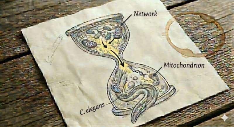
The focus is on phosphatidylcholine—a fundamental lipid that is a major component of biological membranes. It ensures that membranes remain flexible and can dynamically reorganize themselves. Precisely this property is crucial for so-called “mitochondrial fusion”—a process in which individual mitochondria merge into networks. These networks are necessary for cells to distribute key molecules—such as cellular energy equivalents, metabolic products, DNA, and signaling molecules—and facilitate their exchange, thereby preventing imbalances and replacing damaged components.

The study shows that the body’s production of phosphatidylcholine declines with age, leading to increased fragmentation and dysfunction of mitochondrial membranes. When genes involved in phosphatidylcholine synthesis were deactivated in young worms, their mitochondria in the cells quickly began to look “aged.”

The researchers were particularly fascinated by how closely these changes resembled the mitochondria typically observed in chronologically old organisms. Even more striking was the observation that the mitochondria regained a more youthful structure within just two days when the worms were fed phosphatidylcholine or its precursor, choline.

“We were surprised ourselves by how strongly this molecule influences the structure, connectivity, and function of mitochondria,” explains Dr. Tetiana Poliezhaieva, the study’s first author.

‘Butterfly effect’ of a small biochemical change

What initially sounds like a small biochemical change has far-reaching consequences (Butterfly effect). Normally, mitochondria form a dynamic network within the cell, which can constantly adapt to new demands. With age, however, this network becomes increasingly unstable. “You can imagine the whole system as a finely branched power grid that becomes increasingly damaged with age: connections break down and currents stall,” explains Dr. Ermolaeva, the study’s lead author.

“Although energy production continues, it becomes less efficient and sustainable, and energy can no longer be distributed flexibly.” As a result, cells gradually lose their “metabolic plasticity,” meaning their ability to quickly and efficiently adapt to changing energy demands.

This adaptability is essential for maintaining healthy function over time, not only at the level of individual cells but also across tissues and whole-body physiological systems. Its decline is therefore increasingly recognized as a key feature of aging, and it is also closely associated with diseases such as diabetes.

Methodological approach: From worms to humans

To decipher the underlying mechanisms, the research team combined multiple complementary model systems, including the nematode Caenorhabditis elegans, human cell cultures, and large-scale clinical patient data. Using a longitudinal, across-aging approach, they integrated extensive datasets covering proteomic and lipidomic profiles, genetic variation, gene expression, and metabolic activity in humans.

This multi-layered strategy allowed them to connect molecular changes observed in model organisms with patterns seen in human aging. This integrative approach—combined with experimental validation and whole-body functional analyses in worms—made it possible to uncover a direct mechanistic link between gradual molecular changes and systemic aging processes.

New insights into the aging process

The study found that, in addition to accumulating genetic damage, age-related changes in lipid synthesis also contribute to mitochondrial dysfunction. These findings expand our understanding of mitochondrial aging by identifying membrane lipid dynamics as an additional key factor.

A longitudinal comparative study of different life stages of the nematode was also of interest. The data suggests that aging does not proceed uniformly, but rather in phases with different biological breaking points. First, cells lose their capacity to cope with stress, alongside impairments in protein homeostasis—the system that maintains protein stability. This is followed by metabolic and, finally, epigenetic changes.

Sex-specific differences in lipid metabolism were also found: The strongest relative decrease in phosphatidylcholine levels was detected in human metabolome data in women around the age of menopause. “This observation is particularly noteworthy, as it coincides with a time when many women report a significant decline in energy levels and the onset of persistent fatigue,” adds Dr. Ermolaeva.

Aging biology can be modulated

Perhaps the most important finding of the study, however, lies in the reversibility of aging-associated failures: through a targeted increase in phosphatidylcholine levels—for example, via diet—the mitochondrial networks in old C. elegans stabilized, and the cells began producing energy more efficiently again. This indicates that at least some aspects of aging can be substantially restrained, allowing for a longer period of healthy life—and that targeted interventions in metabolism could make a difference.

“Our work shows that both mitochondrial aging and broader systemic aging are, at least in part, modifiable. If we understand the underlying processes, we may be able to take targeted countermeasures,” summarizes Dr. Ermolaeva. Whether and how these findings can be translated into concrete therapies for humans must be clarified in further studies. The role of nutrition is particularly interesting in this context: certain nutrient supplements might help stabilize cell function in old age.

Finally, this study shows that phosphatidylcholine supplementation can serve as an effective anti-aging intervention even when initiated at middle or advanced age. Overall, the study provides an important impetus for aging research. It shifts the focus from irreversible decline to modifiable processes, thereby offering hope that healthy aging can be more actively shaped in the future.