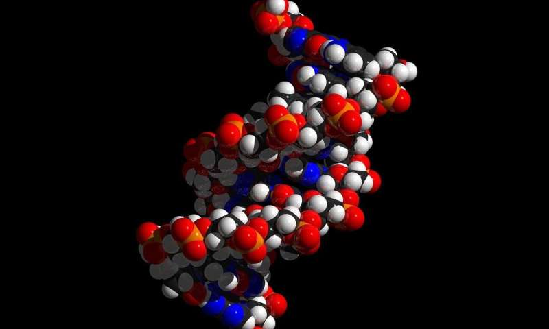
Scientists develop faster technique to uncover hidden gene switches

Researchers at the Max Delbrück Center have developed a new method to discover how DNA controls genes. Their technique, published in Cell Genomics, can reveal the genetic “switches” that regulate important genes more quickly than existing methods.

Most of the human genome does not code for proteins. Instead, much of it consists of regulatory regions. Like switches that turn lights on and off, these regions of nucleotides—called transcriptional enhancers—determine where and when a gene is active, and largely control how much of the corresponding protein a cell produces.

Defects in the genetic code of such regulatory elements can cause developmental defects and disease. But compared to protein-coding regions, they are difficult to identify because they are often located far from the genes they regulate and lack a well-defined genetic code.

Scientists led by Dr. Dubravka Vučićević in the Computational and Regulatory Genomics lab of Professor Uwe Ohler have created a powerful new tool to uncover these regions that control our genes.

Called targeted single-cell activation screen (TESLA-seq), it combines CRISPR-based gene activation (CRISPRa)—a gene regulation technique that uses an engineered form of the CRISPR-Cas9 system to enhance the expression of specific genes—and targeted single-cell RNA sequencing to identify regulatory regions more quickly and accurately than other methods.

“With this method, we can actually test how thousands of candidate regulatory elements in the genome are capable of switching genes on—and find out exactly what genes they influence,” says Vučićević, lead author of the study.

Mapping regulatory elements

To showcase the technique, the study focused on a gene called PHOX2B, which is essential for nervous system development. Mutations in the gene have been linked to neuroblastoma, a cancer of nervous system tissue that primarily affects children.

They concentrated on a large region surrounding PHOX2B, designing 2–3 guide RNAs (gRNAs) for each 100bp segment, or chunk of DNA, for a total of 46,722 gRNAs. This set probed the entire genomic landscape of the PHOX2B gene, as well as other neighboring genes, for potential roles as regulatory switches.

They then transferred each gRNA into a single human neuroblastoma cell. The gRNA told the CRISPRa system where to go and directed it to activate any regulatory regions that might be present in the “chunk.” They identified more than 600 regions—called CaREs (CRISPRa-responsive elements)—that altered cell growth when activated.

The team then zoomed in on about 200 CaREs in more detail and used targeted single-cell RNA sequencing to read out both the gRNA inside each cell and the RNA expressed from nearby genes. This allowed them to link each CaRE to any of the over 70 genes in the PHOX2B region, whose expression changed in that cell. They also found direct connections between CaREs, important regulators of SHISA3 and APBB2, which are involved in cancer and Alzheimer’s disease.

Surprisingly, many CaREs control genes far away, skipping over nearby genes entirely—something other methods often miss.

“TESLA-seq doesn’t just capture what’s happening in one cell type, it can reveal potential connections between genes and regulatory regions across different biological systems,” says Ohler.

This is significant because many diseases affect more than a single tissue type, adds Vučićević.

“The technique can be used to study the vast, uncharted parts of our DNA that influence health and disease across multiple organ systems and can help us to design more precise and effective therapies.”

Molecular biomimetics: The cell nucleus as a model for DNA-based computer chips

In human cells, there are about 20,000 genes on a two-meter DNA strand—finely coiled up in a nucleus about 10 micrometers in diameter. By comparison, this corresponds to a 40-kilometer thread packed into a soccer ball. Despite this cramped space, stem cells manage to find and activate the correct genes in a matter of minutes. Which genes these are differs from cell to cell. Precise activation is crucial as errors in gene selection can lead to disease or cell death.

The investigations of KIT researchers have shown that biomolecular condensates enable fast yet reliable activation of the right genes. “Biomolecular condensates are tiny drops that form in specific places on the DNA—similar to the droplets on the bathroom mirror after a hot shower—and behave like oil in water,” says Professor Lennart Hilbert from the Institute of Biological and Chemical Systems (IBCS) at KIT. “They contain molecular machines, in other words a collection of certain molecules that are necessary for activating genes.”

This process is reminiscent of a key principle in computer science that underlies modern computers and smartphones: the von Neumann architecture. In this architecture, a single processor can very quickly connect to a single address in a large memory, often called RAM. The researchers now want to apply this principle to artificial, DNA-based computer chips to be able to control biotechnological and biomedical applications, for example.

The work is published in the journal Annals of the New York Academy of Sciences.

Surfaces that do the math

“To replicate such biomolecular condensates, in other words the computing centers of the cell nuclei, and build artificial DNA nanostructures for computer chips, we combine traditional lab experiments with modern computer simulations. Using the digital models of DNA nanostructures, we can understand and even predict the behavior of the condensates,” says Mona Wellhäusser, doctoral researcher at IBCS and one of the paper’s co-authors.

To this end, the scientists digitally simulate a system in which enzymes work like small machines and perform specific tasks, like carrying out calculations, for example. To get these enzymes to the right place on the DNA, they use surface condensation, in which the enzymes accumulate by themselves in specific places on the DNA—exactly where they are needed. If candidates that behave correctly are identified in the simulation, they will be synthesized in the lab and examined in test tubes for their actual properties.

“This speeds up the research process enormously, as computer simulations require much less time than lab experiments,” says Hilbert. “So far, we’ve only been able to access one address. But thanks to our research, we’re paving the way for developing a more comprehensive address system and completely new, DNA-based storage and computer systems, the architecture of which is modeled on nature.”

The scientists say that the coronavirus mRNA vaccine and a recently successful patient-specific, “programmed” gene therapy are already demonstrating the potential of biotechnologies that can be programmed by DNA and RNA. Another promising field of application are “DNA chips” for the intelligent control of cancer therapies. They could reprogram immune cells so that they become active as soon as they encounter cancer cells.

Watching how accessory proteins regulate filament growth in real time

Using optical tweezers, researchers at National Taiwan University have observed individual binding events in real time, offering new insights into the molecular regulation of homologous recombination.

Their new study published in Nucleic Acids Research reveals how accessory proteins regulate RAD51 filament growth, a critical step in homologous recombination and DNA repair.

The research, led by Prof. Hung-Wen Li (Department of Chemistry) and Prof. Peter Chi (Institute of Biochemical Sciences), bridges biophysics and biochemistry to address one of the central challenges in DNA repair: how RAD51 assembles on DNA in real time.

Traditional structural techniques capture only static or averaged views of protein clusters, leaving unanswered which oligomeric forms actively drive filament growth.

To overcome this, the team applied single-molecule optical tweezers, which use focused laser beams to manipulate DNA molecules attached to microscopic beads. As RAD51 proteins bound and extended along DNA, the resulting length changes were tracked with nanometer precision, enabling researchers to directly observe stepwise filament growth.

This approach uncovered a striking regulatory role of accessory proteins. RAD51 alone assembled primarily in octameric units, but in the presence of the SWI5-SFR1 complex, the assembly shifted to tetramers. This remodeling stabilized RAD51 filaments, making the assembly process more uniform and efficient.

These insights highlight how accessory proteins fine-tune recombination to safeguard genome integrity. Beyond advancing our understanding of DNA repair, the findings hold implications for cancer biology and genome editing technologies.

“This work demonstrates how single-molecule platforms, coupled with interdisciplinary collaboration, can illuminate fundamental biological processes in unprecedented detail,” says Prof. Hung-Wen Li.

CRISPR study reveals surprising role of Cas9 as guardian of bacterial defense

When scientists discovered how bacteria protect themselves against viral invaders, called phages, in the early 2000s, little did they know they’d stumbled upon a revolutionary tool researchers could use to edit the DNA of living cells.

The system, called CRISPR, is built into bacteria’s genome, and with it, bacteria can “remember” past viral attacks by storing genetic fragments of the invader, called spacers.

In the system’s most well-known version, CRISPR-Cas9, these stored memories are later copied into tiny RNA guides that arm the Cas9 protein to act like a weapon to slice up the invader next time the bacteria encounter a matching phage.

To launch its attack, Cas9 also relies on a short DNA tag called a PAM, as a sign for “can cut here.”

Scientists have modified CRISPR-Cas9 to edit animal and human cells and even develop gene therapies. But a critical piece of the puzzle about how the CRISPR-Cas9 system works has been less clear: How do bacteria form new immune memories?

Setting out to study this mystery, researchers from the lab of Yan Zhang, Ph.D., of the Departments of Biological Chemistry and Microbiology and Immunology at the University of Michigan, have overturned a common assumption about Cas9: that unbound from its usual RNA partners, the “empty” or apo form of Cas9 is functionless. Their study is published in the journal Nature.

Capturing new memory

Until recently, only a handful of genetic studies had shed light on how CRISPR-Cas9 forms immune memories—and these were mostly limited to the Type II-A systems used by the bacteria Streptococcus pyogenes (better known as Group A strep) and Streptococcus thermophilus (often found in yogurt).

In those II-A systems, Cas9 must work together with one of its RNA partners, tracrRNA, to help choose the right spot next to a PAM to capture a new memory.

However, in the Type II-C brand that makes up over 40% of the Cas9 family, the memory acquisition process was less understood.

Zhang’s team, including postdoctoral fellow Xufei Zhou, Ph.D., and Ph.D. student Xin Li, developed a novel model system for studying memory acquisition, using the bacteria Neisseria meningitidis (which can cause meningitis), and infecting it with a phage to see whether memories could be formed in a laboratory setting and then tweaking parts of the immune machinery to see what happened.

“When we started the project, Cas9 was already known as the PAM selector for memory acquisition. What was less clear were the roles of the two RNAs, though we assumed they must be also be somehow important for Cas9 to help bacteria acquire new memories,” said Zhang.

New spacers

Following phage infection, the team used deep sequencing and informatic support provided by colleagues Lydia Freddolino, Ph.D., and Rucheng Diao, Ph.D., of the Departments of Biological Chemistry and Computational Biology and Bioinformatics, to examine the “growing” leader end of the CRISPR array.

There, they saw new spacers derived from the viral genome being added.

Next, they deleted the tracrRNA gene and were shocked to find that the acquisition of spacers was greatly stimulated. Putting tracrRNA back dampened the enhanced acquisition back to normal levels.

A similar pattern emerged with their study of crRNA: without crRNA, Cas9 ramped up acquisition; with crRNA restored, acquisition dropped back down.

“Without the RNAs, we saw that memory formation is highly stimulated to a very robust level,” said Zhang. “This is really surprising for us and, I would say, for the entire field as well—because Cas9 has always been known to need the RNA partners, whether as an immune effector or as a gene editor. This is the first report of a biological function for non-RNA-loaded ’empty’ apoCas9.”

Based on these findings, they proposed that Cas9 acts as a monitor for RNA levels: when CRISPR RNA abundance is low, signaling that the bacterial cell carries short CRISPR and fewer immune memories, apoCas9 forms and can dynamically boost spacer acquisition to protect vulnerable bacteria from infection by quickly building up the memory bank.

Short immune memories

The team further demonstrated three natural conditions when bacteria may experience CRISPR arrays that are too short.

One is at the earliest evolutionary stage, when an array has just been born and holds little—if any—spacer content. At this stage, Cas9 would be without its CRISPR RNA partner and mostly exist in the apo form.

The second and third scenarios both involve the sudden collapsing of a longer CRISPR array into a shorter one, either as a way for bacteria to dump unwanted harmful or autoimmunity memories to acquire new beneficial traits, or as a result of homologous recombination, an event where two similar DNA sequences (a.k.a. CRISPR repeats) swap pieces, erasing intervening CRISPR memories in the process.

This study expands the known functions of Cas9 and fills in a critical missing piece of how CRISPR-Cas9 maintains memory homeostasis.

This dynamic feedback loop offers a new way of thinking about how bacteria safeguard the depth of their immune memory. It may also inspire new ways to design CRISPR-based molecular recording or DNA barcoding tools for research and medicine.

Multifunctional mucus and polydopamine spheres enable targeted, adjustable drug delivery

Researchers at the Technical University of Munich (TUM) have developed hollow microspheres made of mucus and polydopamine using a simple and scalable production method. These tiny spheres are intended to serve as packaging for therapeutic substances, for example in joints or on the oral mucosa. Their properties and mode of action can be adjusted by the choice of materials and are also influenced by the surrounding biological environment.

Oliver Lieleg, professor of biopolymer materials, and his team are harnessing the diverse properties of mucins—the key components of the natural linings such as those found in the oral mucosa or the stomach—to create technological solutions in biomedicine.

Their latest development is a multifunctional microsphere made of mucin and polydopamine. The microsphere is designed to enable a retarded release of molecular cargoes at body sites where adhesion of such drug carriers is otherwise difficult, such as on the oral mucosa or on cartilage.

The work is published in the journal Small.

This good adhesion has been tested on animal tissue and is brought about by the strong adhesive properties of polydopamine. At the same time, mucin adds valuable features: it renders the spheres more adjustable regarding their pore size and allows them to act as a natural lubricant.

“In joints, for example, this could help prevent damage created by joint movements. It may also provide a protective coating on injured tissue in the mouth, another advantage in addition to the microspheres’ function as drug delivery agents,” explains Di Fan, first author of the study.

The hollow spheres are easy to produce, load, and seal

Production of the new hollow spheres starts with an established process: a core is first coated with the desired materials and then removed, leaving behind a hollow structure. With other materials, the spheres sometimes shrink or even collapse when the core is dissolved.

In contrast, the microspheres made from polydopamine and mucin remain structurally stable. Their surface is porous. This allows the cargo to be added after microsphere production and to enter the spheres by diffusion, as shown with model cargo molecules in the study.

The next step is new and crucial: the researchers apply an additional component to the surface, which partially seals the shells of the spheres. This helps keep more of the cargo inside the sphere after loading while ensuring that it is gradually released over time. A range of materials can be used for this locking step, but the approach proved particularly effective with silver ions—positively charged atoms of silver.

Protect or destroy: How material selection and the biological setting determine the effect
The choice of material for sealing is also crucial for the effects of the microspheres. “If silver ions are used, the microspheres help kill cells. This could be particularly useful in treating tumors,” says Di Fan, who demonstrated different effects in cell cultures.

In contrast, without silver ions, the anti-inflammatory properties of polydopamine take effect and protect cells from chemical stress instead. This is especially useful in tissue suffering from inflammation, with potential applications in cases such as osteoarthritis or chronic wounds. Both the type of sealing agent used and the biological setting influence how quickly the cargo is released.

“With the hollow microsphere system, we have created a versatile drug release system that is easy to produce, scalable, and adaptable,” explains project leader Lieleg.

“Our chosen combination of mucin and polydopamine brings together many advantages offered by those biomolecules that go beyond the typical tasks of a classical drug release system; for example, it can protect or eliminate cells—depending on the envisioned application.”

Researchers advance technology for protecting engineered cells

Genetically engineered cell lines used in biomedical research have long been prone to misidentification and unauthorized use, wasting billions of dollars each year and jeopardizing critical scientific discoveries. These problems not only undermine reproducibility of research results, but also put valuable intellectual property at risk.

Now, researchers at The University of Texas at Dallas have developed a novel method to embed unique genetic identifiers in engineered cell lines, eliminating identification errors and safeguarding innovations with tamper-proof genomic tags.

“There are thousands of genetically engineered cell lines in use today, yet we often have no reliable way to verify their identity and origin,” said Dr. Leonidas Bleris, professor of bioengineering in the Erik Jonsson School of Engineering and Computer Science. “Our team has been tackling this challenge by developing innovative solutions that embed unique genetic IDs—essentially barcodes—directly into cells.”

Bleris is corresponding author of a study published in the journal Advanced Science demonstrating the technology.

Custom-designed cell lines are essential for developing vaccines and targeted therapies across a wide range of diseases. The widespread use of the gene-editing tool CRISPR has accelerated the creation of new research models, but this rapid growth has outpaced current authentication capabilities, Bleris said.

“Existing methods can’t reliably distinguish between cell lines that share the same origin but carry different genetic modifications,” Bleris said. “This leaves biomedical research vulnerable due to misidentification, cross-contamination and unauthorized use, and can result in the loss of valuable intellectual property.”

Inspired by a security technology used to protect microchips, UT Dallas researchers have developed a patent-pending method that applies the concept of physical unclonable functions, or PUFs, to living cells—creating unique, tamper-proof genetic “fingerprints” that can’t be copied.

“Biotechnology companies can now ‘barcode’ their cell lines to protect their product,” Bleris said.

In 2022, UT Dallas researchers developed a two-step version of the genetic PUFs technology to protect the authenticity of engineered cell lines. Their new research reduces the process to one step, making the technology easier to implement.

The process uses CRISPR to guide Cas9, an enzyme that acts like a pair of scissors to cut DNA at specific locations. The researchers target the area of the genome called a “safe-harbor” location, where modifications can be made without affecting the cell’s function.

The method leverages another enzyme, terminal deoxynucleotidyl transferase, to repair the break while adding random extra DNA sequences into the safe-harbor area. The added sequences form a unique pattern across the cell population that serves as the unique identifier.

The researchers also developed machine learning tools that can verify cell lines’ identity.

“The machine learning-based method we developed allows us to fully utilize the space of genetic fingerprints and improve the resolution of cell-line identification,” said Taek Kang Ph.D.’23, a bioengineering researcher at UT Dallas, a former Eugene McDermott Graduate Fellow and the study’s co-lead author.

Eel-inspired robots? Study reveals how amphibious animals navigate tough terrain

An international research team has developed an innovative model that explains how elongated amphibious animals—such as eels—coordinate movement both in water and on land. Their study is published in the Proceedings of the National Academy of Sciences.

This collaborative effort, supported by the Human Frontier Science Program, involved researchers from the BioRob lab at EPFL in Switzerland, the Ishiguro Lab at Tohoku University in Japan, and the Standen Lab at University of Ottawa.

Emily Standen, Associate Professor at uOttawa’s Faculty of Science and one of the lead Principal Investigators, led the biological side of the research. “Our study introduces a new model to explain the control of locomotion in elongated amphibious animals,” she says. “We aim to deepen our understanding of the neuromotor control systems used by animals that can adapt their movements between aquatic and terrestrial environments.”

The research, which has spanned multiple years, involved a comprehensive approach combining simulation modeling at Tohoku University, robotics testing at EPFL, and animal observation at the University of Ottawa.

“In my lab, we observed eels to better understand their motor control systems and observe how brain signals, local spinal pattern generators and sensory feedback systems influence undulatory locomotion,” Professor Standen explains. “By using eels as a living model, we were able to guide the simulation and robotics models with biological data.”

The models in this study show that basic components of the motor system, like coordination in the nervous system, as well as pressure feedback and stretch feedback, allow for redundant coordination during swimming. This redundancy and the capacity of stretch feedback to allow the exploitation of heterogeneity in the environment to help move forward, may explain why elongated fish like the eel and lamprey can move in terrestrial environments.

“These animals are remarkably resilient,” she notes. “Our models point to sensory feedback as the key to allowing them to maintain their locomotor performance.”

Bio-inspired robotics

Beyond animal biology, the findings could help engineers design flexible robots for challenging environments. “This research provides new ways of understanding neuromotor control in animals, which can have far-reaching implications for both scientific research and technological advancements,” says Professor Standen. Imagine robots that crawl, slither, or swim through tight spaces, using nature’s engineering to stay flexible and strong.

The study, titled “Multisensory feedback makes swimming circuits robust against spinal transection and enables terrestrial crawling in elongate fish,” is a leap forward in understanding movement and could inspire innovative robot designs in the future.

Engineered E. coli produce biodegradable plastic that outperforms widely used PET

The PET (polyethylene terephthalate)-alternative PDCA (pyridinedicarboxylic acid) is biodegradable and has superior physical properties, according to a recent study. A Kobe University team of bioengineers engineered E. coli bacteria to produce the compound from glucose at unprecedented levels and without byproducts—and opened up a realm of possibilities for the future of bioengineering. The findings are published in the journal Metabolic Engineering.

The durability of plastics is both the reason why they have become so widespread and why they pose environmental problems. In addition, they are mainly sourced from petroleum, making them nonrenewable and contingent on geopolitics. Research groups worldwide work on both biodegradable and bio-sourced alternatives, but there often are issues with yield, purity and—as a result—associated production cost.

Kobe University bioengineer Tanaka Tsutomu says, “Most biomass-based production strategies focus on molecules consisting of carbon, oxygen and hydrogen. However, there are highly promising compounds for high-performance plastics that include other elements such as nitrogen, but there are no efficient bioproduction strategies. And purely chemical reactions inevitably generate unwanted byproducts.”

PDCA is such a candidate. It is biodegradable, and materials incorporating this show physical properties comparable to or even surpassing those of PET, which is widely used in containers and textiles.

“Our group approached the challenge from a new angle: We aimed to harness cellular metabolism to assimilate nitrogen and build the compound from start to finish,” says Tanaka.

The Kobe University group has now achieved the production of PDCA in bioreactors at concentrations more than seven-fold higher than previously reported.

Tanaka explains, “The significance of our work lies in demonstrating that metabolic reactions can be used to incorporate nitrogen without producing unwanted byproducts, thereby enabling the clean and efficient synthesis of the target compound.”

The group, however, did have some stubborn problems to solve along the way. The most stubborn of these came when they discovered a bottleneck where one of the enzymes they had introduced produced the highly reactive compound hydrogen peroxide, H2O2. The compound then attacked the enzyme that produced it, thereby deactivating it.

“Through refining the culture conditions, in particular by adding a compound that can scavenge H2O2, we could finally overcome the issue, although this addition may present new economic and logistical challenges for large-scale production,” says Tanaka.

The bioengineers already have plans on how to improve the production going forward, with every problem pointing the way to its solution. Looking at the future, Tanaka says, “The ability to obtain sufficient quantities in bioreactors lays the groundwork for the next steps toward practical implementation. More generally, though, our achievement in incorporating enzymes from nitrogen metabolism broadens the spectrum of molecules accessible through microbial synthesis, thus enhancing the potential of bio-manufacturing even further.”

AI turns printer into a partner in tissue engineering

Organ donors can save lives, for example, those of patients with kidney failure. Unfortunately, there are too few donors, and the waiting lists are long. 3D bioprinting of (parts of) organs may offer a solution to this shortage in the future. But printing living tissues, bioprinting, is extremely complex and challenging.

The team of Riccardo Levato at UMC Utrecht and Utrecht University is now taking an important step toward printing implantable tissues. Using computer vision, a branch of artificial intelligence (AI), they’ve developed a 3D printer that doesn’t just print, it also sees and even co-designs.

Their research was published today in Nature. With this innovation, they tackle one of the biggest challenges in 3D bioprinting: improving both the survival and functionality of cells in printed living tissue. But how exactly does that work?

We usually associate 3D printing with building structures layer by layer. But there are other forms, such as volumetric bioprinting. This technique creates a complete structure in a single step, using a light-sensitive gel that solidifies when exposed to cell-friendly laser light. The advantage? It is incredibly fast, taking just seconds, and much gentler on living cells.

To produce a high-quality print, it is crucial to understand what’s inside the printing material, so that the printed object is built as optimally as possible. The new technology, called GRACE, makes that possible. It opens up new possibilities for bioprinting functional tissues, and brings us closer to repairing tissues, testing new drugs, and even replacing entire organs.

What is 3D-bioprinting?

In 3D bioprinting, researchers use living cells to create functional tissues and organs. Instead of printing with plastic, they print with living cells. This comes with great challenges. Cells are fragile and wouldn’t survive a regular 3D printing process. That’s why Levato’s team developed a special bio-ink, a mix of living cells and nourishing gels that protect the cells during the printing process.

With the advancements in bio-inks, layer-by-layer 3D bioprinting became possible. But this method is still time-consuming and puts a lot of stress on the cells. Researchers from Utrecht came up with a solution: volumetric bioprinting.

Volumetric bioprinting is faster and gentler on cells. Using cell-friendly laser light, a 3D structure is created all at once. “To build a structure, we project a series of light patterns into a spinning tube filled with light-sensitive gel and cells,” Levato explains. “Where the light beams converge, the material solidifies. This creates a full 3D object in one go, without having to touch the cells.” To do this, it is crucial to know exactly where the cells are in the gel. GRACE now makes that possible.

Sammy Florczak, a Ph.D. student in Riccardo’s lab, worked on the development of GRACE, short for Generative, Adaptive, Context-Aware 3D printing. He built a new device in a specialized lab, using advanced laser technologies. Before entering, a red light signaling “LASER” shows whether it’s safe to go in.

Laser light plays a crucial role, not just in the printing step, but also in the added imaging step that sets this new technology apart. GRACE combines volumetric bioprinting with this advanced laser-based light-sheet imaging. But what can we do with that?

Smart blood vessels around living cells

One of the biggest challenges in 3D bioprinting is creating functional blood vessels. Blood vessels are essential to provide oxygen and nutrients to the cells, and thus printing these blood vessels in the correct place is key to creating viable tissues.

Yet, in conventional printing methods, a 3D design is made before knowing where the cells are located in the light sensitive gel and thus where the blood vessels must be printed. With GRACE, the printer “sees” where the cells are located and, within seconds, designs a network of blood vessels around those cells as effectively as possible.

“In the past, printing always depended on the designer’s blueprint. Now, GRACE contributes to the design itself,” Florczak explains. “The printer ‘sees’ what kind of cells are in the material, and where they are. Then, using AI tools, it creates a matching design for the object to be printed. This new printer essentially has its own ‘eyes’ (the laser-based imaging) and ‘brain’ (the new AI software). That level of customization leads to tissues that survive and function better.”

GRACE can do more than create adaptive blood vessel networks. The technology can also align multiple printing steps automatically. Take a piece of printed bone tissue, for example, that later needs a layer of cartilage added. Normally, that is a complex process with a lot of manual work. GRACE scans the existing tissue and automatically designs and prints a second layer that fits perfectly on top. All at the high printing speed of volumetric bioprinting, creating cm3-sized objects within seconds.

Another challenge in bioprinting is that light can sometimes be blocked, for example by previously printed parts of the structure. This can create shadows and flaws in the final product. GRACE can solve this too. By scanning the surface of any obstacles, the system automatically adjusts the light projection. This makes the print more precise and consistent. Moreover, this allows pre-made objects to be inserted into the printing vial. Think, for example, of a stent in which you could print blood vessel cells or objects that can release medicines.

Just the beginning

Bioprinting is highly promising, but significant work is still needed to translate this technology to the clinic. Riccardo underlines that further research is needed to determine how printed cells can mature to replicate the functionality of native tissues. Even considering the challenges ahead, Riccardo is not afraid to dream big.

“This first work on GRACE is just the beginning. We are now working on increasing the amount of cells that can be printed, so that other tissues like heart and liver can also be printed. Moreover, we would like to make this technique openly accessible to other labs, so other could apply it to their printing method.”

Mapping the lipid blueprint of vertebrate life in 4D

Researchers at EPFL have created the first 4D lipid atlas of vertebrate development, revealing how fats shape our bodies from embryo to organism.

We often think of embryonic development as a genetic ballet, choreographed entirely by DNA and proteins. But there’s another cast member quietly shaping the scene: lipids. These fat molecules aren’t just fuel; they play structural, signaling, and even patterning roles as embryos develop.

Despite advances in genomics, we still don’t fully understand how metabolism is arranged in different parts of the body during development. A big part of the metabolism puzzle are lipids, which vary widely in structure and function and have been notoriously hard to map across entire organisms both in high resolution and across time.

Previous techniques only offered fragmented snapshots, but without a detailed atlas, scientists can’t track where and when specific lipids appear in the developing body. This has limited our ability to understand not just basic biology, but also how metabolic disorders or congenital diseases might take root.

A team of researchers at EPFL have developed a new computational method that allowed them to build the first 4D lipid map of a vertebrate embryo—specifically, the zebrafish. “4D” refers to mapping lipids in the three dimensions of space plus the fourth dimension of time, which captures how lipid distributions change as the embryo develops. Using an innovative combination of imaging mass spectrometry and a new computational framework called uMAIA, they tracked more than 100 lipid types across space and time.

The research was led by Professors Gioele La Manno and Giovanni D’Angelo, working with the group of Andrew Oates. Their study is published in Nature Methods.

The team used a technique called MALDI mass spectrometry imaging to scan “slices” of zebrafish embryos at different stages of development, allowing them to measure where different lipids are located in tissue slices. But processing this kind of data is not easy, as each zebrafish embryo generates huge amounts of spectrometry data, making an impossible puzzle.

To handle the data, the team developed uMAIA (“unified Mass Imaging Analyzer”), a powerful algorithm that extracts, aligns, and normalizes the data into coherent, accurate maps. It basically, turns a pile of noisy data into a clear movie of metabolic development. uMAIA applies adaptive image extraction, matching similar molecules across sections, and correcting technical noise. What emerges is a detailed, high-resolution atlas of how lipid distribution changes from early embryo to full-fledged fish.

The team found that lipids form highly organized patterns that match anatomical structures. For example, certain sphingolipids—which are important for cell membranes and signaling—accumulated in the swim bladder, a fish organ that is analogous to human lungs. Others concentrated in developing brain regions or bone-forming areas. These spatial patterns suggest lipids play key roles in shaping organ function and identity.

Knowing where and when lipids appear can help researchers understand developmental diseases, like congenital metabolic disorders. It could also inform regenerative medicine or tissue engineering. And because lipid metabolism is often disrupted in diseases like cancer or Alzheimer’s, this atlas offers a baseline for comparison.

“From this effort emerges not only a powerful resource but a Swiss army knife for doing this kind of mapping again and again across other systems in health and disease,” says Prof. La Manno.