Ultra-processed foods tied to nearly fourfold asthma risk in children

Children who get more than 30% of their daily energy from ultra-processed foods (UPFs), such as soda drinks, packaged snacks, and sweetened breakfast cereals, have a nearly fourfold risk of developing asthma in their early school years.

The finding comes from a new study published in the journal Allergy that followed nearly 700 Spanish children for an average of 3.4 years as part of the SENDO (Seguimiento del Niño para un Desarrollo Óptimo) project.

The diet-asthma connection

Asthma is a common, long-term condition that affects breathing and often runs in families. While genetics are known to play a role, environment and lifestyle factors are also to blame. In this study, researchers wanted to know whether certain parts of our modern diet, specifically ultra-processed foods, were associated with the later development of asthma and other allergic diseases.

They followed 691 children who were 4 or 5 years old when the study started. Parents reported what their children ate by completing a detailed questionnaire, and the team categorized the foods using the NOVA system. This is a framework that classifies foods by processing levels rather than nutrient content.

Parents also updated the researchers each year on whether their children had been diagnosed with asthma or allergies. To ensure accuracy, the team took into account numerous factors that might skew the results, such as the child’s weight and how much time they spent in front of screens.

A clear pattern emerges

The results showed a strong link between ultra-processed foods and asthma. “Higher UPF consumption may be associated with an increased risk of developing asthma in school-age children,” commented the team in their paper.

Beyond the nearly fourfold increase in asthma risk compared to children with the lowest intake of UPFs, the study found that the quantity of processed foods mattered. As the percentage of processed food in a child’s diet went up, the likelihood of an asthma diagnosis also appeared to increase.

However, no such link was shown between ultra-processed foods and other allergic conditions. The study authors suggest that this may be because UPFs are irritating the lungs through a type of inflammation not related to an allergic reaction.

Given the link between diet and asthma suggested in the study, the scientists issued a call to action. “These findings highlight the need for public health policies aimed at limiting processed food consumption in children’s diets as a preventive strategy.”

Viagra could hold key to halting Peyronie’s disease

Combining two widely prescribed drug classes could provide the first effective treatment for early-stage Peyronie’s disease, according to a new study published in The Journal of Sexual Medicine.

Peyronie’s disease (PD) is caused by the development of fibrotic scar tissue within the penis, leading to pain, curvature, sexual dysfunction and, in many cases, significant psychological distress. It affects an estimated 10% of men during their lifetime, but despite its prevalence, treatment options are limited, particularly in the early phase of the condition.

The study, carried out by Anglia Ruskin University (ARU) and University College London Hospital (UCLH), found that combining phosphodiesterase type 5 (PDE5) inhibitors such as sildenafil (Viagra) and tadalafil (Cialis) with selective estrogen receptor modulators (SERMs), including tamoxifen, may slow or even stop disease progression when given early.

The clinical study, carried out by Professor David Ralph of UCLH, evaluated outcomes in 133 men diagnosed with acute Peyronie’s disease who were treated with the drug combination for three months. Their results were compared with a smaller group of patients receiving standard care, which included giving vitamin E or no treatment at all. Standard care did not include surgery.

The study found 43% of patients on the combination experienced an improvement in penile curvature, almost three times higher than in the standard-care group (15%).

At the start of treatment, 65% of patients in the combination group reported pain during erections. After three months, that figure had fallen to just 1.5%. By comparison, pain prevalence in the standard-care group fell from 50% to 27%.

The clinical findings build on earlier laboratory work led by Professor Selim Cellek at ARU’s Fibrosis Research Group.

Over the course of several years, Professor Cellek’s team screened 1,953 FDA-approved drugs to identify compounds capable of blocking the transformation of fibroblasts into myofibroblasts, the key cells responsible for fibrosis.

PDE5 inhibitors and SERMs emerged as particularly effective, and when used together demonstrated an effect greater than either drug alone.

Currently, there are no approved oral therapies proven to prevent early disease progression, forcing patients in the acute phase to wait until the condition stabilizes before they can be offered treatments including injections or surgery.

Professor Cellek said, “Positive findings from this pilot clinical study validate our drug-screening approach in the lab. It shows how repurposing well-known medicines can accelerate progress in areas of unmet clinical need.

“Because both PDE5 inhibitors and SERMs are already widely used in clinical practice and have established safety profiles, the approach could be readily adoptable if confirmed in larger studies.

“These results suggest that early intervention targeting fibrosis could change how we treat Peyronie’s disease. Repurposing existing drugs may allow us to move from managing symptoms to modifying the disease itself.”

Professor David Ralph, Professor of Urology at UCLH, said, “This paper confirms the basic science research with regards to halting the progression of Peyronie’s disease. In previous papers we have noted that tamoxifen and PDE5 inhibitors inhibit the transformation of fibroblasts into myofibroblasts and therefore contraction of the plaque.

“This has now been put into clinical practice, where this paper shows that when tamoxifen and a PDE5 inhibitor are combined, there is statistically less progression of the disease and improvement in curvature compared to the control arm. This is where from bench to clinical practice prevails and, hopefully now, a prospective clinical trial can be initiated.”

Diseases can spread between apartments via shared ventilation, study shows

Airborne diseases like measles, influenza and COVID-19 can easily spread between units in multi-family buildings via a type of bathroom ventilation system commonly used around the world, new research suggests. The study, conducted inside an older high-rise in Spain early in the coronavirus pandemic, adds to a growing body of evidence that airborne viruses can spread between separated indoor spaces, transmitting disease without face-to-face contact.

“We tend to think that if we shut the door in our apartment, we are safe and can’t get infected. But our study shows that, depending on the ventilation system in place, that may not be the case,” said senior author Shelly Miller, professor emerita in the Department of Mechanical Engineering at the University of Colorado Boulder.

The findings were published in the journal PLOS One on May 12.

‘The ghost in the room’

The project got its start in Santander, Spain, in June of 2020, just as residents began to emerge from lockdown. Cases had dropped to zero, and people were still masking and social distancing, when residents of a seven-story building got a call from public health officials. Someone on the third floor had tested positive for the SARS CoV2-virus.

The building was locked down again. But soon, 15 people across four vertically stacked apartments had contracted COVID-19.

Engineer David Higuera, who lived in the building, had a hunch that the ventilation shafts connecting bathrooms were to blame.

“I knew that if what my wife and I suspected was happening, it could have significant scientific implications for public health,” said Higuera, a co-author on the study.

Higuera partnered with Miller, a world expert in airborne aerosols who was on sabbatical in Spain, and engineers and epidemiologists from the University of Valencia and the University of Cantabria in Spain, and Concordia University in Canada.

They genetically sequenced samples from sickened residents, confirming that infected individuals likely got it from one another. Then they measured airflow and air pressure in the building. In one experiment, the team remotely measured carbon dioxide (CO2) levels throughout the day in a vacant apartment.

CO2 is emitted through human breath and wouldn’t be expected to exist at high levels in a vacant space. Yet the apartment was filled with it.

“It was like there was a ghost in the room,” said Higuera.

As with many older buildings across Spain and around the world, the bathrooms weren’t designed to have windows or fans. Instead, they use the “stack effect” in which shared vertical ventilation shafts move air out through vents in the wall, exhausting it through the roof via natural convection.

The study found that shifts in weather could impact air pressure in the shaft, with hotter temperatures pushing airflow back through the vents. Switching on the kitchen exhaust hood could exacerbate the spread, sucking almost all the air from an adjacent bathroom into a nearby apartment within minutes.

“The most plausible transmission route for this outbreak was the bathroom vertical ventilation duct system,” the authors conclude.

Could it happen elsewhere?

In 2003, during an outbreak of severe acute respiratory syndrome (SARS) in a Hong Kong high-rise, virus-laden aerosols made their way into a shared ventilation system via a bathroom floor drain, infecting 321 and killing 42 people.

The Santander building was built in 1969, and that type of ventilation system was phased out in Spain in 1975 with new building codes. But roughly one-third of the country’s buildings were built before then.

This type of ventilation system is uncommon in the United States, but similar systems still exist worldwide in older buildings.

“While this is a special building design more common in Spain, it illustrates a broader concern—that even if you are far from the source, if your air is connected, you can still get sick,” said Miller. “This can happen in a multifamily apartment building through the ducts, in a hotel between the hallway and rooms off the hallway, in office buildings between offices or on a cruise ship.”

Higuera installed a fan in his own bathroom during the pandemic, with a flap that prevented airflow from coming in. No one in his family got sick.

The authors are calling on officials in Spain to update building standards to assure that older buildings take similar steps to mitigate disease transmission risk. They say building designers and managers everywhere should do more to address indoor air quality.

Breast cancer cells with doubled genomes may dodge immunotherapy by turning off key immune signals

An epigenetic mechanism by which tumors manage to hide from the body’s immune defenses has just been described by an international scientific team led by the University of Liège and the Dana-Farber Cancer Institute. These findings open up avenues for new therapeutic strategies combining epigenetics and immunotherapy. The paper is published in the journal Cancer Cell.

Whole-genome doubling (WGD) is an event that frequently occurs during tumor development; it is observed in approximately 37% of primary solid tumors (tumors that form a physical, compact mass within an organ or tissue) and in up to 56% of metastatic tumors (secondary tumors that have developed after cancer cells have broken away from the primary tumor and migrated to other organs via the blood or lymphatic circulation). This duplication corresponds to a situation where the cancer cell ends up with twice the normal number of chromosomes. Long considered merely a marker of poor prognosis, this phenomenon was associated with resistance to treatment and increased genomic instability, without its mechanisms of action being clearly understood.

A new international study sheds significant light on how this mechanism develops, particularly in breast cancer. The researchers show that duplication does not merely disrupt the structure of the genome; it actively alters the relationship between the tumor and the immune system.

“Initially, genomic duplication increases the tumor cell’s visibility to the immune system,” explains Dr. Pierre Foidart, medical oncologist at the Liège University Hospital (CHU de Liège) and researcher at the University of Liège (fellowship of Foundation Against Cancer), and first author of the article. “But the cells quickly adapt by developing a strategy of invisibility.”

Normally, a cancer cell displays on its surface fragments of abnormal proteins called antigens. These antigens are presented by a structure known as the major histocompatibility complex class I (MHC-I), which acts like a “display window” allowing cytotoxic CD8+ T lymphocytes – immune cells specialized in destroying abnormal cells – to recognize and eliminate the cancer cell.

At the same time, certain cells of the innate immune system, particularly NK cells, produce a signaling molecule called interferon gamma (IFN-γ). This cytokine increases the amount of MHC-I present on the surface of cancer cells, thereby improving antigen presentation to CD8+ T lymphocytes.

Once activated, CD8+ T lymphocytes also produce interferon gamma, further enhancing the recognition of tumor cells. This creates a positive feedback loop that progressively amplifies the anti-tumor immune response.

“What our study demonstrates is that genome-doubled cells eventually switch off the genes responsible for MHC-I production, and therefore for antigen presentation,” continues Pierre Foidart. “They no longer display antigenic fragments on their surface and no longer respond correctly to interferon signals. T lymphocytes, deprived of the information they need to recognize the threat, pass by the tumor without launching an attack.” The cancer cell is still there, however, but it has become, in a sense, invisible to the immune system.

This camouflage is not genetic but epigenetic (the set of mechanisms that regulate genes without altering the DNA sequence). The DNA remains intact, but its expression is modulated by metabolic changes that lead to transcriptional repression of immune genes.

“This mechanism is driven in part by the PRC2 complex, an epigenetic regulator. And this is precisely what distinguishes epigenetic modifications from genetic mutations: epigenetic modifications are reversible, unlike genetic mutations,” says Dr. Kornélia Polyak, Professor of Medicine at Dana-Farber Cancer Institute, Harvard Medical School, and a co-leader of the Dana-Farber Harvard Cancer Center Cancer Cell Biology Program.

“By pharmacologically targeting this complex, it is possible to partially restore antigen presentation and make WGD-positive tumor cells recognizable to the immune system once again, while selectively inhibiting the growth of WGD-positive tumors.”

These findings have potential implications for clinical practice. Whole-genome duplication could eventually serve as a biomarker for stratifying patients and guiding treatment decisions, particularly towards combinations of epigenetic inhibitors and immunotherapy. Clinical studies will, however, be necessary to validate these approaches.

Pierre Foidart is currently continuing his research on this topic in collaboration with Professor Pierre Close (ULiège), with the aim of developing methods for detecting whole-genome duplication that are more accessible than whole-genome sequencing, which remains costly and is rarely used in routine clinical practice.

New study challenges the idea that testosterone drives risk-taking behavior

Men are more likely to take risks in tricky situations than women, but whether there is an inherent biological reason behind it is a question researchers have been asking for quite some time. A popular theory suggests that higher levels of the hormone testosterone are what make men less risk-averse. A recent study, however, debunked this notion.

A review spanning dozens of studies involving more than 17,000 participants found no reliable link between testosterone and how much risk a person chooses to take. Rather than being driven by a single hormone, risk-taking seems to stem from a mix of biological, psychological, and social factors. A separate meta-analysis looking at sex differences found that testosterone’s link to risk-taking behavior is no stronger in men than in women. The findings are published in Neuroscience and Behavioral Reviews.

Testing the testosterone theory

Testosterone is the main male sex hormone and plays a key role in male development. It is primarily produced in the testicles and helps drive changes during puberty, such as a deeper voice, facial and body hair growth, and increased muscle strength. Although men naturally have higher levels of testosterone, women also produce it in their gonads, the ovaries. Both men and women need testosterone to maintain bone strength, energy levels, muscle growth, and healthy red blood cell production.

Many researchers have zeroed in on testosterone as a key driver of gender differences in risk aversion—a person’s tendency to choose safer options with predictable outcomes rather than riskier choices that may offer greater rewards but involve uncertainty. This behavior shapes decisions both large and small, from how fast we drive, where we invest our money, and what activities we seek out on holiday, to high-stakes choices in politics, international relations, and economic policy.

Given the far-reaching impact of risk-taking and aversion, scientists have spent years trying to decode this complex aspect of human behavior. While some pointed to social and cultural factors, others focused on the potential biological driver. However, the evidence on testosterone has been very inconsistent. Some studies linked higher testosterone to greater risk-taking, while others found no relationship at all, and some even found the opposite.

To make sense of the existing literature on this topic, the researchers conducted a meta-analysis of 52 studies involving 17,340 participants that examined both testosterone and risk-taking. The studies they included used a wide range of testosterone measurement methods, including direct approaches such as blood and saliva tests, as well as hormone administration. Across the included studies, risk-taking was assessed using gambling tasks, lottery games, and personality questionnaires.

A statistical method called multilevel random-effects meta-analysis was used to pool results across studies, accounting for differences in study design and the fact that many studies included multiple measurements. In addition, the researchers conducted a separate analysis to determine whether the findings differed between men and women or by task type.

Even though men naturally have higher testosterone and often take more risks than women, testosterone does not explain this difference in behavior, as there was no clear association between the hormone and the behavior. The researchers noticed that a tiny link only appeared in studies that used lottery-style games, whereas other risk-taking measures showed no meaningful relationship. They also found that studies using direct measures of testosterone often reported no link between the hormone and risk-taking levels, whereas studies using indirect methods, such as finger-length ratios (an indicator of prenatal testosterone exposure), were more likely to suggest a possible association.

The results make it quite evident that risk-taking is not driven by hormones alone. The researchers suggest that risk-taking likely depends on a biopsychosocial framework, involving a combination of the situation at hand, how a person thinks and feels, and the specific demands of the task. The debate is likely to remain open until future studies use larger sample sizes, standardized hormone testing methods, and more consistent measures of risk-taking.

Digital aging twin measures how organs age at different speeds across adulthood

Aging is a complex process, and precisely measuring how the human body declines has long been a challenge. Two people of the same chronological age can have very different health trajectories. Scientists have also struggled to move beyond identifying aging markers to pinpointing what actually drives aging itself.

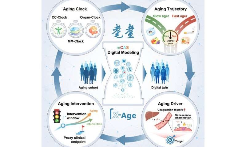
Now researchers from China’s Aging Biomarker Consortium (ABC) have built a computational framework—the Digital Aging Twin—to study aging at the individual level in order to predict biological age and track the different aging rates of individual organs.

The study, which was conducted by researchers from the Institute of Zoology, the China National Center for Bioinformation (Chinese Academy of Sciences), Xuanwu Hospital of Capital Medical University, and seven other institutions, marks a major breakthrough by moving from simply describing aging to systematically quantifying it, potentially paving the way for future interventions.

The findings were published in Cell on May 8.

Building a massive aging dataset

The team recruited 2,019 healthy individuals aged 18 to 91 from Chinese cities including Beijing, Quzhou, Ningbo, and Nanchang to create a standardized multicenter cohort called mCAS (multicentric Chinese Aging Standardized). The researchers collected data on 240 parameters for each participant, using clinical tests, cognitive and motor function assessments, brain and retinal imaging, gait analysis, and several layers of molecular data, including DNA methylation, RNA transcripts, proteins, metabolites, and gut microbiomes.

This dataset, comprising more than a billion high-quality data points, serves as the foundation for a three-tiered system of “clocks” to measure aging.

How the three-tier clock works

The first tier is the core capacity clock, which integrates 240 physiological indicators to reflect overall functional decline. The second and most powerful tier is the multimodal clock. It integrates multiple layers of molecular data (“omics” layers) via a deep learning process that employs “attention mechanisms”—which prioritize the most informative data—to quantify the contribution of different data types. This multimodal clock predicts chronological age with a mean absolute error of only 3.87 years, outperforming all single-omics clocks. The third tier comprises organ-specific clocks for the brain, liver, lungs, muscles, blood vessels, and skin, each based on clinical markers, plasma proteins, and imaging features.

One of the most striking findings is that organs age asynchronously. For example, the liver reaches a critical aging inflection point around age 40, whereas the brain’s aging accelerates at around age 50. The analysis also uncovered two major nonlinear waves of aging-related change: one occurring between ages 40 and 50, and another between 60 and 70.

Pinpointing coagulation factors as drivers

Seeking the causes of these aging-related changes, the researchers analyzed plasma proteomics, examined stained liver tissue from human donors, and conducted experiments using human cell cultures and animal models. They identified age-driven accumulation of liver-derived coagulation factors—particularly F13B, as well as F9 and F10—as a direct driver of vascular and systemic aging.

For example, when human aortic endothelial cells were exposed to these factors, they showed clear signs of senescence: elevated aging markers, impaired tube formation (a measure of blood vessel health), and increased inflammation. Similarly, injecting F13B into mice accelerated aging across multiple tissues, including the liver, heart, aorta, and kidney, accompanied by immune cell infiltration and inflammatory signals. These results show that coagulation factors are not just passive biomarkers but actionable drivers of aging.

Simplifying clocks for clinical use

To make the aging clock approach clinically practical, the researchers developed simplified “proxy clocks” using just 100 to 108 plasma proteins. These protein-based proxies closely match the predictions of the much more complex core capacity clock and organ clocks, suggesting that a relatively simple blood test might one day provide a comprehensive aging assessment.

The study also identified lifestyle factors influencing biological aging. Greater fruit intake, consistent sleep routines, and moderate walking were linked to slower aging. In contrast, smoking, insufficient sleep, and high meal frequency were associated with accelerated aging.

Implications for China’s X-Age Project

This research marks the first proof-of-concept achievement of the X-Age Project (also known as “耄耋”), a major national initiative led by the Aging Biomarker Consortium to build a comprehensive system of aging clocks for the Chinese population.

Although the current aging-clock framework is built on cross-sectional data, it is being continuously refined with longitudinal follow-up data and larger, more diverse populations. Future updates will also generate lower-cost and more sensitive detection approaches.

Despite the limitations of using cross-sectional data, the study has far-reaching implications for aging research as a whole. The Digital Aging Twin framework represents a fundamental shift in aging science—from description to prediction and from identifying correlations to pinpointing drivers.

Researchers now have a standardized, quantifiable, and interpretable system that can tell how fast a person is aging, which organs are aging most rapidly, and where interventions might be most effective.

RNA therapy slows harmful heart remodeling after heart attack in clinical trial

Following an acute heart attack, pathological remodeling processes occur in the heart. One consequence is so-called left ventricular systolic dysfunction, in which the pumping function of the left ventricle is impaired. To compensate for this, the heart muscle enlarges excessively, thereby becoming further weakened. The key regulator of this harmful growth of heart muscle cells is microRNA-132 (miR-132).

A team led by Prof. Dr. Thomas Thum, Director of the Institute for Molecular and Translational Therapeutic Strategies at Hannover Medical School (MHH), has produced a synthetic antagonist called CDR132L, which can block the main switch for cardiac hypertrophy and reverse chronic heart failure. The researchers have already demonstrated this in animal models and early clinical trials.

The drug candidate CDR132L has now been investigated in an international Phase II clinical trial involving patients who have recently suffered a heart attack and have heart failure. The HF-REVERT study has shown that patients with already advanced cardiac remodeling at the start of the study could benefit particularly from treatment with CDR132L. The results of the study were published in the journal Nature Medicine.

MicroRNA regulates pathological heart muscle growth

MicroRNA molecules belong to a class of molecules known as non-coding RNA (ncRNA), meaning they are not translated into specific proteins. Instead, they regulate a wide range of cellular processes—such as how a cell grows, whether it divides, or what type of cell it develops into. On the other hand, excessive microRNA activity can alter gene regulation and thereby trigger diseases.

One example of this is miR-132. More than ten years ago, Professor Thum’s research team discovered that a massive accumulation of this microRNA is directly linked to the pathological proliferation of heart muscle cells. The antisense oligonucleotide blocker CDR132L is the first ncRNA-based therapy to be used in Phase II trials for heart disease.

CDR132L is particularly beneficial for seriously ill patients

The HF-REVERT trial was conducted at around 80 study centers across seven European countries and the United Kingdom. A total of 294 patients were randomly assigned to three groups within three to 14 days of suffering a heart attack. In addition to standard treatment for heart failure, they received either CDR132L in two different doses or a placebo, administered in three intravenous doses at four-week intervals. The analysis was based on 280 patients who had received at least one dose.

“Our study has demonstrated that CDR132L is safe and well-tolerated and does not cause any harmful side effects on the liver, kidneys, the hematopoietic system or the heart,” says Professor Thum. CDR132L was particularly effective in patients with already advanced cardiac remodeling, meaning their hearts were already severely damaged.

“These findings support the further clinical development of the drug, particularly in the field of chronic heart failure,” emphasizes the cardiologist. The results represent an important step towards RNA-based therapies in cardiology. They offer great potential for positively influencing the progression of the disease in patients with heart failure.

CDR132L was developed by Cardior Pharmaceuticals GmbH, which was founded in 2016 by Prof. Dr. Dr. Thomas Thum and is based on his research at the Institute for Molecular and Translational Therapy Strategies. This MHH spin-off highlights the importance of university-based innovation for the development of new therapeutic approaches. Two further clinical trials are currently underway in patients with chronic heart failure, led by the Danish pharmaceutical group Novo Nordisk, which acquired the biopharmaceutical company Cardior in 2024.

Like mother, like fetus: Study finds contagious yawning begins in the womb

Yawning is incredibly contagious, and more often than not, seeing someone yawn right in front of us makes us instinctively do the same. It is often tied to social and emotional connection and brain mirroring, where we automatically align and simulate the emotions and actions of the people around us. A recent study published in Current Biology has found that this behavior begins even before birth.

Researchers recorded the facial expressions of pregnant women while an ultrasound machine captured real-time images of their fetuses’ faces. By comparing the two recordings, the researchers observed that fetuses were more likely to yawn after their mothers yawned, with a delay of about 90 seconds.

Tracking mother–fetus yawn sync

Yawning in humans begins far earlier than most people realize. Fetuses start yawning in the womb at around 11 weeks of development. Since there is no air for the fetus to draw in, during a yawn, they slowly open their mouths, perform movements that resemble breathing in and out, and then gently close their mouths again. For a long time, scientists believed that fetal yawning was thought to be driven purely by internal biological processes, but there wasn’t enough evidence to prove it either right or wrong.

In this study, the researchers wanted to see if fetuses in the womb would catch a yawn from their mothers. For this, they recruited 38 pregnant women who were between 28 and 32 weeks along, all with healthy, uncomplicated pregnancies.

The experiments involved the mothers watching three different types of video in a quiet room: a yawning video, a mouth-movement video, and a still-face video. While a video camera monitored the mother’s face, the researchers used a 2D ultrasound machine to provide a real-time view of the fetus’s nose and lips.

Three experts, who didn’t know what the mother was watching, reviewed the collected footage and verified the yawns. The researchers used an AI tool called DeepLabCut to precisely track subtle lip and nose movements, then trained a neural network to see whether a mother’s yawn mirrored the movement pattern of her fetus’s.

The researchers found that fetal yawning increased significantly only when the mother yawned, not when she simply opened and closed her mouth or kept her face still. They called this phenomenon prenatal behavioral contagion. The fetal yawns were not random either; they typically appeared about 90 seconds after the mother yawned, which is similar to the response time seen in contagious yawning among adults.

These findings suggest that fetal yawning may be part of an early mother-baby connection, where a mother’s behavior can influence how the fetus responds. Further research into how deeply this behavioral connection works, and whether it has long-term developmental effects, could reshape prenatal care.

Treatment-resistant depression may yield to combinations of medications already in clinical use

Many people with major depressive disorder get no relief from current treatments. Newer combinations of existing medications might help, researchers report in JAMA Psychiatry.

Persistent low mood, a loss of interest in previously enjoyable activities, lack of energy, feelings of worthlessness, poor concentration and appetite, and suicidal thoughts are symptoms of major depression. A significant percentage of adults suffering from it don’t get much relief from conventional antidepressant therapies. Doctors then have to look for alternative therapies to help these patients.

“At least one-third of adults with depression do not respond to at least two trials of conventional antidepressant therapies. These patients are considered to have treatment-resistant depression, and alternative therapies should be considered for such patients,” says UConn School of Medicine psychiatric epidemiologist T. Greg Rhee.

Rhee and colleagues at Harvard, Yale, and the University of Toronto, among others, have two recent studies in JAMA Psychiatry evaluating existing drugs used in new ways to treat major depression. One looks at the efficacy of intravenous ketamine, and the other at combinations of antidepressants with anti-psychotics.

Ketamine was originally developed as a fast-acting surgical anesthetic. There is also evidence it can rapidly alleviate depression in some individuals. The US Food and Drug Administration has approved esketamine, a version of the ketamine molecule, in nasal spray form as a treatment for depression. But intravenous ketamine is still being evaluated.

The researchers analyzed 26 existing randomized controlled trial studies that compared intravenous ketamine with controls. They found that ketamine was more effective than a placebo over the short term of a few days, but the effects were less pronounced after a few weeks. And ketamine seemed to work about as well as esketamine. Both drugs were very effective in rapidly reducing suicidal impulses in people who were in immediate danger of harming themselves.

Their second JAMA Psychiatry study compared how well combinations of antidepressants with antipsychotics worked in people with treatment-resistant depression. The researchers performed a meta-analysis of 22 studies, looking both at reduction of depressive symptoms and at side effects of the drugs. They found some antipsychotics were significantly more likely to help decrease symptoms of depression. But the antipsychotic that was most likely to help, lumapeterone, was also most likely to be discontinued by the patient because of side effects.

“These studies could potentially guide practicing psychiatrists and other clinicians to consider these new approaches of modalities for patients with moderate to severe depression, who did not previously respond to conventional antidepressant therapies,” says Rhee.

Rhee also adds, “We plan to conduct population-level epidemiologic studies to further examine the effectiveness and safety profiles of these treatment options.”

Gold-coated microneedles can detect subtleties in how liver and kidneys process drugs in real time

Scientists have taken a giant leap forward with the development of tiny microneedles designed to detect subtle but critical changes in how the liver and kidneys process therapeutic drugs. The experimental technology, under development at the University of California, Los Angeles, aims to overcome longstanding limitations that have hindered wearable microneedle biosensors.

“Wearable microneedle biosensors promise real-time molecular monitoring for precision medicine but are limited by low sensitivity and tissue abrasion,” writes Dr. Jialun Zhu, lead author of a new study published in Science Translational Medicine.

“Overcoming these challenges, we recast electrode functionality not merely as a sensing substrate but as a mechanism for resilient, high signal-to-noise ratio measurements in tissue,” added Zhu, a bioengineer in UCLA’s Samueli School of Engineering.

In plainer English, the multidisciplinary UCLA team was able to get their system to work flawlessly compared with similar devices by other research groups.

Tracking drug clearance in real time

The team has developed a biosensor that in early research already shows promise for real-time in-tissue monitoring of drug pharmacokinetics. These preclinical studies also show that the device is both safe and highly accurate. However, it has not yet been tested in humans.

Still, if the technology seems futuristic, it may be because tiny wearables and implants that measure any number of biological processes have been themes in science fiction for decades. With refinements underway to improve precision medicine, the future has already arrived. Scientists involved in the project ranged from molecular and cellular biologists to biochemists and a team of bioengineers like Zhu.

One goal in precision medicine has been the development of a minimally invasive device that can monitor the clearance of drugs from a patient’s kidneys and liver, providing more accurate dosing guidelines. To address persistent problems that have impeded progress toward reaching that goal, the team engineered what it calls a “resilient nanostructured bioelectrode” using a microscopically thin layer of a precious metal.

“Our microneedle-based resilient nanostructured bioelectrode is fabricated using a bilayer process that strengthens the electrode with a micrometer-thick gold adhesion layer,” Zhu noted in the research paper.

The key reason that an accurate wearable biosensor is needed is explained by the growing number of medications with narrow therapeutic ranges. That means it is possible to provide doses that are either too low or too high. With a wearable such as the one under development, doctors can tell if they have prescribed a precise dose, and how well the drug is being processed and excreted.

Toward precision monitoring in organ dysfunction

In preclinical experiments, the biosensor enabled continuous in-tissue monitoring of drug pharmacokinetics, including changes associated with liver and kidney dysfunction. Scientists found in the animal model research that their experimental technology measured drug kinetics for six days and produced accurate parameters for drug dosing while also monitoring drug clearance from the liver and kidneys.

The system revealed, for example, that one chemotherapy drug, irinotecan, cleared out slowly in mice with liver damage. The technology also traced the kinetics of several antibiotics during various stages of chronic kidney disease.Comparison between conventional blood-based therapeutic drug monitoring and wearable ISF-based therapeutic drug and metabolic function monitoring. Credit: Science Translational Medicine (2026). DOI: 10.1126/scitranslmed.adr5493

An approach by other research groups has involved the use of wearable biosensors that incorporate microneedles, which measure minute molecular changes in drug concentrations. However, current microneedles suffer from issues such as low sensitivity and poor mechanical durability.

In contrast, Zhu and colleagues developed a more resilient, nanostructured microneedle that analyzes the biochemistry of interstitial fluids between cells. Their design incorporates sensors that endow it with a high degree of specificity and features a strong layer of gold that increases the needle surface area and resists corrosion.

Technology holds promise

In an editorial commentary, Molly Ogle, an associate editor at Science Translational Medicine, notes that wearable technology could play an important role in precision medicine. “The study demonstrates preclinical promise for minimally invasive therapeutic drug monitoring and functional assessment of hepatic and renal drug processing,” Ogle wrote.

Zhu and colleagues underscored, meanwhile, that their device not only has marked improvements over similar technology but also could be economically manufactured. They predict that their resilient nanostructured bioelectrode could be mass produced at less than $1.50 per sensor.

“These results establish the resilient nanostructured bioelectrode as a viable microneedle platform for high-fidelity in vivo deployment of electrochemical biosensors, enabling minimally invasive, longitudinal monitoring of low-concentration analytes and real-time assessment of organ function,” Zhu and the UCLA team concluded.